. 2009 Mar;119(3):661-73.
doi: 10.1172/JCI37515. Epub 2009 Feb 23.

Confirming the RNAi-mediated mechanism of action of siRNA-based cancer therapeutics in mice

Affiliations

Confirming the RNAi-mediated mechanism of action of siRNA-based cancer therapeutics in mice

Adam D Judge et al. J Clin Invest. 2009 Mar.

Abstract

siRNAs that specifically silence the expression of cancer-related genes offer a therapeutic approach in oncology. However, it remains critical to determine the true mechanism of their therapeutic effects. Here, we describe the preclinical development of chemically modified siRNA targeting the essential cell-cycle proteins polo-like kinase 1 (PLK1) and kinesin spindle protein (KSP) in mice. siRNA formulated in stable nucleic acid lipid particles (SNALP) displayed potent antitumor efficacy in both hepatic and subcutaneous tumor models. This was correlated with target gene silencing following a single intravenous administration that was sufficient to cause extensive mitotic disruption and tumor cell apoptosis. Our siRNA formulations induced no measurable immune response, minimizing the potential for nonspecific effects. Additionally, RNAi-specific mRNA cleavage products were found in tumor cells, and their presence correlated with the duration of target mRNA silencing. Histological biomarkers confirmed that RNAi-mediated gene silencing effectively inhibited the target's biological activity. This report supports an RNAi-mediated mechanism of action for siRNA antitumor effects, suggesting a new methodology for targeting other key genes in cancer development with siRNA-based therapeutics.


Figures

Figure 1
Figure 1. Activity of PLK1 siRNAs in vitro.
Correlation between mRNA silencing and HT29 cell viability for (A) PLK1424 (P1424), (B) PLK773 (P773), and (C) LUC siRNA treatments. PLK1 mRNA was determined by bDNA analysis at 24 hours. Duplicate plates were assessed for cell viability at 72 hours. (D) PLK1424 siRNA causes dose-dependent reductions in viability of LS174T, HT29, Hep3B, and HepG2 cell cultures. Cells were treated for 72 hours with PLK1424 SNALP at increasing concentrations of 0.3, 0.6, 1.25, 2.5, and 5 nM siRNA (white bars to black bars, respectively). Values in AD are expressed as percentage of viability or PLK1 mRNA relative to PBS control and reflect mean of 3 separate experiments (± SD) conducted in triplicate cultures. (E) Decreased cell viability is associated with the induction of apoptosis. Caspase-3/7 activity in LS174T cells was assessed 24 hours and 48 hours after treatment with SNALP-encapsulated PLK773, PLK1424, and LUC control siRNAs. Data represent fold induction over PBS in triplicate cultures (mean ± SD triplicate cultures).
Figure 2
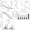
Figure 2. In vitro activity of unmodified versus 2′OMe-modified PLK1 and KSP siRNA.
Activity of the 2′OMe-modified panels of (A) PLK1424 and (B) PLK773 siRNA. Unmodified PLK1424 or PLK773 siRNA was compared in the Hep3B cell viability assay with the 2′OMe-modified duplexes 1/A, 2/A, 1/B, 2/B, 1/C, and 2/C that comprise the respective 2′OMe sense/AS oligonucleotides (see Table 1). Data show mean viability of triplicate cultures relative to PBS-treated cells and represent 2 independent experiments using SNALP-formulated siRNAs. (C) Cytokine induction by unmodified and 2′OMe PLK1 siRNA in vitro. Murine Flt3L DCs were treated with 5 μg/ml (350 nM) unmodified PLK773 and PLK1424 siRNA duplexes and their constituent sense (S) or AS oligonucleotides or the 2′OMe siRNA duplexes PLK773-1/B (1/B) and PLK1424-2/A (2/A) formulated in SNALP. IFN-α and IL-6 were assayed in culture supernatants at 24 hours. Values represent mean + SD of 3 separate experiments conducted in triplicate cultures. (D and E) Activity of SNALP-formulated KSP2263 siRNA in murine Neuro2a cells. (D) Correlation between KSP mRNA silencing and cell viability relative to PBS control. KSP mRNA was determined by bDNA analysis at 24 hours. Duplicate plates were assessed for cell viability at 72 hours. (E) Activity screen comparing the unmodified KSP2263 siRNA to KSP2263-U/U (U/U), KSP2263-G/U (G/U), and KS2263-G/G (G/G) siRNA duplexes that comprise the respective sense/AS 2′OMe oligonucleotides (see Table 1). SNALP-formulated KSP2263 siRNA were tested in the Neuro2a cell viability assay. Data represent mean ± SD triplicate cultures, relative to PBS treatment.
Figure 3
Figure 3. 2′OMe-modified PLK1, KSP, and LUC siRNA induce no measurable IFN response in mice.
SNALP-formulated LUC (unmodified) and 2′OMe-modified LUC-U/U, PLK1424-2/A, PLK773-1/B, and KSP2263-U/U (K2263 U/U) siRNA were administered at 2 mg/kg i.v. to BALB/c mice. (A) IFIT1 relative to GAPDH mRNA in liver and spleen was assessed at 4 hours by bDNA analysis. (B). Serum IFN-α was assessed at 6 hours by ELISA. SNALP-formulated 2′OMe siRNAs induced no detectable increase in either IFN-α protein or IFIT1 mRNA relative to PBS vehicle. Values represent mean + SD (n = 4).
Figure 4
Figure 4. Therapeutic activity of PLK1 and KSP siRNA in hepatic tumors.
PLK1424-2/A treatment confers significant survival advantages in SCID/beige mice bearing hepatic Hep3B tumors. Mice were administered SNALP-formulated PLK1424-2/A (n = 15) or LUC-U/U (n = 8) at 6 × 2 mg/kg i.v. twice weekly (day 10 to day 28). (A) Body weights (mean + SD) over the dosing period expressed as percentage of initial weight on day 10. (B) Kaplan-Meier plot of days to euthanization due to tumor burden. PLK1424-2/A treatment provided significant survival advantage over control treatment. (P = 0.03, log-rank Cox-Mantel test). (C) Residual hepatic Hep3B tumor burden in mice 24 hours after final administration of PLK1424-2/A siRNA (5 × 2 mg/kg siRNA on days 8, 11, 14, 18, and 21). Bars represent hGAPDH mRNA/mg liver of individual mice (mean ± SD of triplicate analyses) determined by human-specific bDNA assay. No tumor, livers from non–tumor-seeded mice. See Supplemental Figure 6 for additional data. (D) KSP2263-U/U treatment confers survival advantages in A/J mice bearing hepatic Neuro2a tumors. Mice were administered SNALP-formulated KSP2263-U/U or LUC-U/U (n = 8) at 5 × 4 mg/kg i.v. (q3d ×5 from day 8 to day 21 after tumor seeding). Kaplan-Meier plot of days to euthanization due to tumor burden. End points are based on clinical scores as a humane surrogate for survival. Mean SNALP particle sizes were 83 (0.09 polydispersity), and 90 (0.12 polydispersity) nm for PLK1424-2/A and LUC-U/U formulations, respectively.
Figure 5
Figure 5. Target mRNA silencing in hepatic tumors by the RNAi mechanism.
(A and B) Target mRNA silencing and (C and D) detection of RNAi-specific mRNA cleavage products in tumors following SNALP-formulated siRNA administration. SCID/beige mice with established intrahepatic Hep3B tumors were administered a single 2 mg/kg dose of SNALP-formulated PLK1424-2/A or LUC-U/U siRNA, and RNAi activity was assessed by (A) PLK1 mRNA in tumor lysates and (C) 5′ RACE-PCR analysis. (A) Tumor (human) PLK1/GAPDH mRNA ratios 24 hours after siRNA administration (mean ± SD of 4 animals). (C) RACE-PCR detects the specific 5′ cleavage product of PLK1 mRNA from tumors analyzed in A. Lanes represent PCR products derived from individual PBS (n = 2), LUC-U/U (n = 2), and PLK1424-2/A–treated mice (n = 3). (B) Mouse KSP mRNA and (D) 5′ RACE-PCR analysis of resected hepatic Neuro2a tumors from mice treated with SNALP-formulated KSP2263-U/U siRNA. Data are presented as in A and C. Positive control from in vitro Neuro2a cell lysates treated with KSP2263-U/U siRNA indicated by plus sign; no template control indicated by minus sign. RACE-PCR detects the specific 5′ cleavage product of mouse KSP mRNA from tumors. Identities of the predicted 476-bp PLK1 and 102-bp KSP PCR products (arrows) were confirmed by direct DNA sequencing. Mean SNALP particle sizes were 83 (0.09 polydispersity), 90 (0.12 polydispersity), and 88 (0.07 polydispersity) nm for PLK1424-2/A, LUC-U/U, and KSP2263-U/U formulations, respectively.
Figure 6
Figure 6. Duration of RNAi activity within hepatic tumors.
(A) 5′ RACE-PCR analysis of Hep3B liver tumors 24 hours, 48 hours, 96 hours, 7 days, and 10 days after a single i.v. administration of SNALP-formulated PLK1424-2/A siRNA (2 mg/kg). Specificity of the PLK1424-specific RACE-PCR product (arrow) was confirmed by sequencing at day 1 and day 7. (B) Corresponding levels of PLK1 mRNA in isolated tumor RNA analyzed in A. Duration of RNAi correlated with duration of mRNA silencing compared with that of LUC-U/U SNALP–treated mice. Data represent mean hPLK1/ hGAPDH mRNA ratio + SD (n = 3 at each time point). Mean SNALP particle sizes were 83 (0.09 polydispersity) and 90 (0.12 polydispersity) nm for PLK1424-2/A and LUC-U/U, respectively.
Figure 7
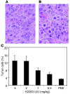
Figure 7. KSP2263-U/U induces distinct phenotypic changes typical of KSP inhibition in hepatic tumor cells.
Hepatic Neuro2a tumor histology 24 hours after a single i.v. administration of (A) LUC-U/U or (B) KSP2263-U/U siRNA formulated in SNALP (2 mg/kg siRNA). Images representative of tumors from at least 6 individual mice. H&E staining reveals tumor cells with aberrant nuclear figures typical of monoastral spindles or apoptotic phenotypes in KSP2263-U/U–treated mice. Original magnification, ×200. (C) Quantitative histology of H&E-stained tumor tissues from mice treated with SNALP-formulated KSP2263-U/U at 4, 2, 1, or 0.5 mg/kg siRNA. Tumor cells with condensed chromatin structures were scored as positive, and the number of such tumor cells was calculated as a percentage of total tumor cells taken from 10 fields of view. Positive cells included aberrant and typical mitotic and apoptotic figures. Values are mean + SD of 3 mice. Mean SNALP particle sizes were 88 (0.07 polydispersity) and 82 (0.08 polydispersity) nm for KSP2263-U/U and LUC-U/U, respectively.
Figure 8
Figure 8. PLK1424-2A induces distinct phenotypic changes typical of PLK1 inhibition in hepatic tumor cells.
H&E tumor histology 24 hours after single i.v. administration of 2 mg/kg SNALP-formulated (A and C) LUC-U/U or (B and D) PLK1424-2/A siRNA. Images are representative of tumors from at least 7 individual mice. Original magnification, ×200 (A and B); ×400 (C and D). Mean SNALP particle sizes were 72 (0.04 polydispersity) and 72 (0.02 polydispersity) nm for PLK1424-2/A and LUC-U/U, respectively.
Figure 9
Figure 9. Therapeutic activity of PLK1 SNALP containing either C14 or C18 PEG-lipids in s.c. tumors.
(A) Inhibition of s.c. tumor growth by alternate PLK1424-2/A SNALP formulations. Mice were administered PLK1424-2/A SNALP comprising either PEG-cDMA or PEG-cDSA (6 × 2 mg/kg i.v.) between day 10 and day 21 after Hep3B tumor seeding. Values show mean tumor volumes (mm3) ± SD (n = 5). Control was LUC-U/U siRNA SNALP (PEG-cDMA). (B) Corresponding hPLK1/hGAPDH mRNA ratio in s.c. Hep3B tumors following single administration (2 mg/kg) of PLK1424-2/A or LUC-U/U siRNA; mean + SD (n = 4). (C) Dose response of PLK1424-2/A PEG-cDSA SNALP in Hep3B tumors. Mice bearing established (~100 mm3) tumors were administered PLK1424-2/A PEG-cDSA SNALP (6 × 3, 6 × 1, or 6 × 0.5 mg/kg), LUC PEG-cDSA SNALP (6 × 3 mg/kg), or PBS vehicle every 2–3 days between days 18 and 29 after seeding. Values represent mean tumor volumes (mm3) (n = 5). Mean SNALP particle sizes were 81 (0.10 polydispersity), 71 (0.03 polydispersity), 82 (0.12 polydispersity), and 74 (0.05 polydispersity) nm for PLK1424-2/A PEG-cDMA, PEG-cDSA, LUC-U/U PEG-cDMA, and PEG-cDSA, respectively.

Comment in

Similar articles

Cited by

References

    1. Elbashir S.M., et al. Duplexes of 21-nucleotide RNAs mediate RNA interference in cultured mammalian cells. Nature. 2001;411:494–498. doi: 10.1038/35078107. - DOI - PubMed
    1. Judge A.D., et al. Sequence-dependent stimulation of the mammalian innate immune response by synthetic siRNA. Nat. Biotechnol. 2005;23:457–462. doi: 10.1038/nbt1081. - DOI - PubMed
    1. Hornung V., et al. Sequence-specific potent induction of IFN-alpha by short interfering RNA in plasmacytoid dendritic cells through TLR7. Nat. Med. 2005;11:263–270. doi: 10.1038/nm1191. - DOI - PubMed
    1. Kleinman M.E., et al. Sequence and target-independent angiogenesis suppression by siRNA via TLR3. Nature. 2008;452:591–597. doi: 10.1038/nature06765. - DOI - PMC - PubMed
    1. Hornung V., et al. 5′-Triphosphate RNA is the ligand for RIG-I. Science. 2006;314:994–997. doi: 10.1126/science.1132505. - DOI - PubMed

Publication types

MeSH terms