. 2009;4(3):341-55.
doi: 10.1038/nprot.2008.250.

A protocol for the production of recombinant spider silk-like proteins for artificial fiber spinning

Affiliations

A protocol for the production of recombinant spider silk-like proteins for artificial fiber spinning

Florence Teulé et al. Nat Protoc. 2009.

Abstract

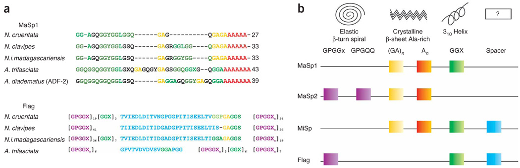
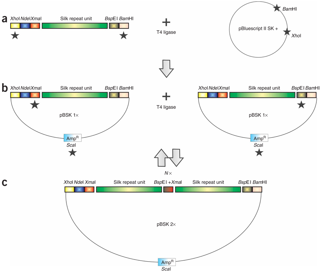
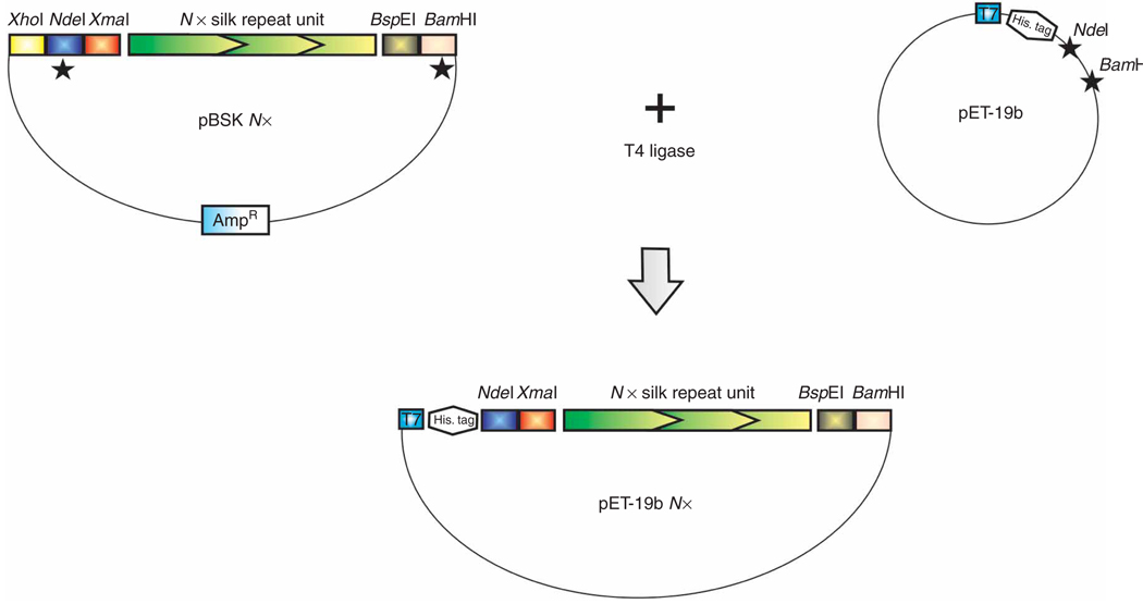
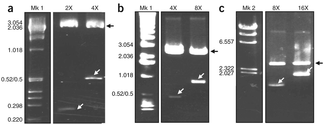
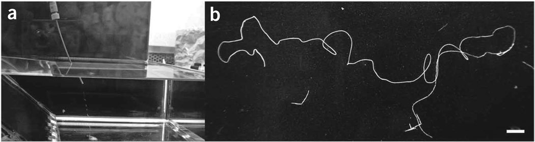
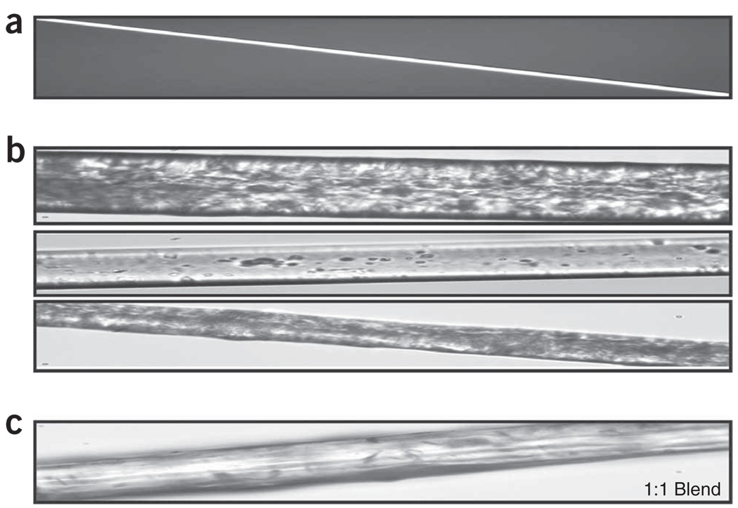
The extreme strength and elasticity of spider silks originate from the modular nature of their repetitive proteins. To exploit such materials and mimic spider silks, comprehensive strategies to produce and spin recombinant fibrous proteins are necessary. This protocol describes silk gene design and cloning, protein expression in bacteria, recombinant protein purification and fiber formation. With an improved gene construction and cloning scheme, this technique is adaptable for the production of any repetitive fibrous proteins, and ensures the exact reproduction of native repeat sequences, analogs or chimeric versions. The proteins are solubilized in 1,1,1,3,3,3-hexafluoro-2-propanol (HFIP) at 25-30% (wt/vol) for extrusion into fibers. This protocol, routinely used to spin single micrometer-size fibers from several recombinant silk-like proteins from different spider species, is a powerful tool to generate protein libraries with corresponding fibers for structure-function relationship investigations in protein-based biomaterials. This protocol may be completed in 40 d.


Figures

Figure 1
Figure 1
The molecular structure of orb-weaver spiders’ silk proteins. Alignment of the consensus repetitive sequences of (a) major ampullate (MaSp 1) and flagelliform (Flag) silk proteins (adapted from ref. 20). Structural amino-acid motifs found consensus repeats of spider silk proteins (b). The square-colored boxes indicate that the structural motif is part of the silk protein. To determine which silk proteins contain which modules, read the line that connects the boxes from left to right, starting with the silk name. The empty box marked ’?’ indicates that the secondary structures of these ’spacer’ motifs are unknown. (Adapted from ref. .) MaSp1 or MaSp2: major ampullate spidroin 1 or 2; MiSp: minor ampullate spidroin; Flag: flagelliform protein.
Figure 2
Figure 2
Strategy to build large synthetic spider silk-like tandem repeat sequences from small double-stranded monomer DNAs flanked by compatible but nonregenerable restriction sites. (a) The engineered silk-like module with appropriate flanking restriction sites is cloned in the plasmid vector. (b) The recombinant plasmid is subjected to two separate restriction digestions and, in both cases, fragments containing the insert are isolated and ligated to each other. (c) The resulting plasmid contains an insert that was doubled in size and has a nonfunctional internal XmaI/BspEI hybrid site. The black stars (★) indicate the restriction digestion of DNA and N× means that the strategy can be repeated as many times as needed.
Figure 3
Figure 3
Strategy to clone the engineered synthetic silk-like sequences in the pET-19b expression vector. The black stars (★) indicate the restriction digestion of DNA.
Figure 4
Figure 4
Agarose gel analyses showing the synthetic Flag/MaSp 2 silk DNA multimers at different doubling stages. The sequential recombinant plasmids containing the different silk-like insert fragments were subjected to restriction digestion with XmaI and BspEI to release the silk insert. The restriction digestion products were separated on (a) Nusieve/agarose 3:1 and (b,c) 0.8–1% agarose gels. After staining with ethidium bromide, the DNA fragments were visualized using UV light. In a–c, Mk: molecular marker; Mk 1:1 kbp DNA Ladder; Mk 2: Lambda DNA-HindIII; 2×−16×: repetitive synthetic silk sequences after sequential insert doubling (i.e., in a, 4× is twice the size of 2×). The size of the silk inserts are 236, 472, 944 and 1,888 bp for 2×, 4×, 8× and 16×, respectively. The black arrows show the linearized pBluescript plasmid and the white arrows show the silk inserts. The molecular weights in kbp are indicated.
Figure 5
Figure 5
Synthetic silk fiber formation by extrusion. The pure lyophilized silk recombinant protein was solubilized in 100% HFIP. The silk spinning dope was loaded in the spinneret constituted of a glass syringe attached to PEEK tubing. Manual extrusion of the dope into a 90% isopropanol coagulation bath (a) generated a uniform silk fiber (b). The photograph in b was taken using a Nikon Coolpix 950 digital camera. The white bar in b represents 1 cm.
Figure 6
Figure 6
Western blot analysis showing the IMAC purification steps of a chimeric Flag/MaSp 2 recombinant protein. The 60-kDa His-tagged A4S88 recombinant protein was detected using the 6× His mAb–HRP conjugate. Mk: molecular weight marker Precision Plus Protein Standard Dual color; F1–6: IMAC collected fractions—F1: unbound proteins; F2–4: wash 1 (20 mM imidazole), wash 2 (40 mM imidazole), wash 3 (50 mM imidazole), respectively; F5: elution fraction (250 mM imidazole); F6: strip fraction. The recombinant protein is highly concentrated in the elution fraction (F5) and residual in the strip fraction (F6). The numbers indicate the molecular weights in kDa.
Figure 7
Figure 7
Native and synthetic spider silk fibers. (a) Native N. clavipes major ampullate fiber has a diameter of 4 µm. (b) MaSp 2-like synthetic spider silk fibers. Notice the highly variable appearance and much larger diameter compared with native fibers. (c) Synthetic spider silk blend produced by extrusion of a 15% (wt/vol) MaSp 1/MaSp 2 spinning dope. These photos were taken using a Nikon Eclipse E200 microscope at ×40 original magnification.

References

    1. Peters HM. Über den spinnapparat von Nephila madagascariensis (Radnetzspinnen Argiopidae) Z. Naturforsch. 1955;10:395–404.
    1. Lucas F. Spiders and their silks. Discovery. 1964;25:20–26.
    1. Foelix RF. Spider webs. In: Foelix RF, editor. Biology of Spiders. 2nd edn. New York, USA: Oxford University Press Inc. & Georg Thieme Verlag; 1996. pp. 110–149.
    1. Akai H. The structure and ultrastructure of the silk gland. Experientia. 1983;39:443–449.
    1. Sehnal F, Akai H. Insect silk glands: their types, development and function, and effects of environmental factors and morphogenetic hormones on them. Int. J. Insect. Morphol. Embryol. 1990;19:79–132.

Publication types