. 2009 Mar 18;28(6):755-65.
doi: 10.1038/emboj.2009.26. Epub 2009 Feb 19.

GTPase activation of elongation factor EF-Tu by the ribosome during decoding

Affiliations

GTPase activation of elongation factor EF-Tu by the ribosome during decoding

Jan-Christian Schuette et al. EMBO J. .

Abstract

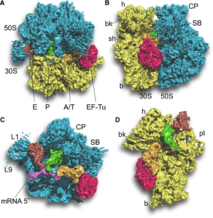
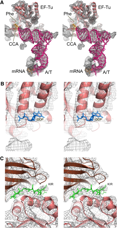
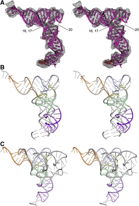
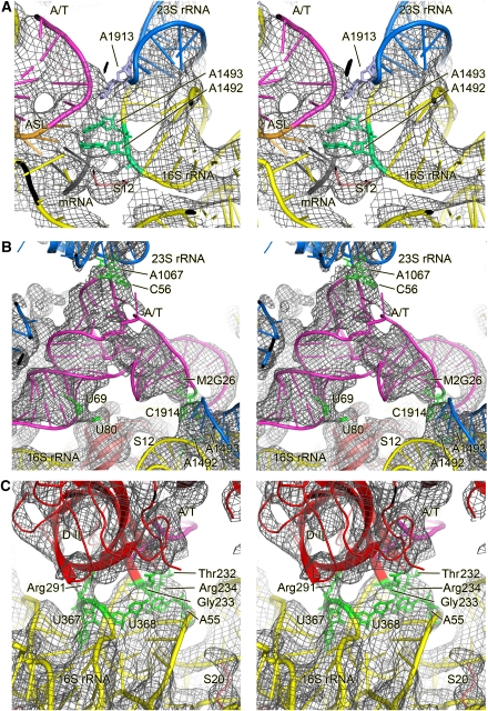
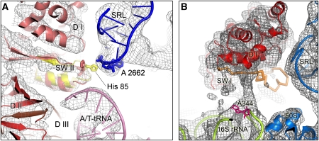
We have used single-particle reconstruction in cryo-electron microscopy to determine a structure of the Thermus thermophilus ribosome in which the ternary complex of elongation factor Tu (EF-Tu), tRNA and guanine nucleotide has been trapped on the ribosome using the antibiotic kirromycin. This represents the state in the decoding process just after codon recognition by tRNA and the resulting GTP hydrolysis by EF-Tu, but before the release of EF-Tu from the ribosome. Progress in sample purification and image processing made it possible to reach a resolution of 6.4 A. Secondary structure elements in tRNA, EF-Tu and the ribosome, and even GDP and kirromycin, could all be visualized directly. The structure reveals a complex conformational rearrangement of the tRNA in the A/T state and the interactions with the functionally important switch regions of EF-Tu crucial to GTP hydrolysis. Thus, the structure provides insights into the molecular mechanism of signalling codon recognition from the decoding centre of the 30S subunit to the GTPase centre of EF-Tu.


Figures

Figure 1
Figure 1
Overview of the 70S•EF-Tu•Phe-tRNA•GDP•kirromycin complex. A surface representation of the cryo-EM map is shown (A) from the top; (B) from the L7/L12 side; (C) from the 30S side, with 30S removed and (D) from the 50S side, with 50S removed. The components are coloured distinctly (30S subunit, yellow; 50S subunit, blue; EF-Tu, red; A/T-tRNA, orange; P-tRNA, green; E-tRNA, brown; mRNA, pink).
Figure 2
Figure 2
Details seen in the electron density map at 5.7- to 6.4-Å resolution. (A) Overall structure of the ternary complex showing the interactions between the EF-Tu and A/T tRNA. The density for the ternary complex has been computationally separated using a mask generous enough to show the sites of interaction with the ribosome. At this resolution, secondary structure elements of RNA and protein are clearly distinguishable. (B) A region of EF-Tu showing additional density that corresponds to GDP. (C) Density for the low-molecular weight kirromycin seen between the domains of EF-Tu.
Figure 3
Figure 3
Distortions in the tRNA during decoding. (A) A molecular model for the A/T-tRNA derived by fitting various segments of the tRNA separately into the electron density. The region of disorder in the D loop is highlighted (green circle, missing nucleotides are labelled) and is consistent with fluorescence changes in a reporter in this loop (see text for details). (B, C) Superposition of the distorted A/T-tRNA (coloured ribbon: acceptor stem, orange; T stem and T loop, cyan; D stem with variable loop, green; D loop, yellow; anticodon stem loop, magenta) and canonical tRNA (grey ribbon). The superpositions were carried out using the acceptor arm (B) and the anticodon stem loop (C), and show that in addition to the kink between the D stem and anticodon stem loop, there is also a rotation of the D loop and stem relative to the acceptor arm.
Figure 4
Figure 4
Structures of the ternary complex on and off the ribosome. Superposition of the molecular model for the ribosome-bound ternary complex (red ribbons) with (A) the X-ray structure of the kirromycin-containing ternary complex (PDB identifier 1OB2, green ribbons) or (B) the X-ray structure of the ternary complex (Nissen et al, 1995) (turquoise ribbons). The ternary complexes are aligned at domain I (G domain) of EF-Tu.
Figure 5
Figure 5
Interactions of the ribosome-bound ternary complex with elements of the ribosome. Stereo representation of the molecular model for the ternary complex and the interacting ribosomal elements (coloured ribbon) docked into the cryo-EM density (grey mesh). Important components or residues are labelled. (A) Details of the decoding centre showing that A1492 and A1493 of 16S RNA are extended out of helix 44 and making contact with the minor groove of the codon–anticodon helix along with G530. Also shown is the base of A1913 in helix 69 of 23S RNA, which is inserted between 16S and 23S RNA and appears to contact the tRNA around position 38 (Selmer et al, 2006). (B) Interactions of the A/T-tRNA with the ribosome outside the decoding region. Residues that are putatively involved in the interactions are highlighted in green. (C) Interaction of the shoulder of the 30S subunit with domain II of EF-Tu. As in (B), residues that are putatively involved in the interactions are highlighted in green. Colour code: blue, 50S subunit; yellow, 30S subunit; pink, A/T-site tRNA.
Figure 6
Figure 6
Interactions of the switch regions of EF-Tu with the ribosome. (A) Close up on the switch II region (SW II) and the sarcin-ricin loop (SRL). The cryo-EM map is shown as grey mesh and models for the docked components as coloured ribbons. In addition, the structure of SW II from the EF-Tu•GMPPNP X-ray structure (PDB identifier 1EXM) is superposed (yellow ribbons) after the structure has been aligned onto the fitted EF-Tu•GDP•aurodox structure (red ribbons), which in turn was docked as a rigid body into the density map. Furthermore, the functionally important His85 (arrow) is highlighted in the docked X-ray structures of EF-Tu•GMPPNP and EF-Tu•GDP•aurodox (we note that we cannot observe the residue directly at the present resolution in the cryo-EM map), as well as A2662 of the SRL (pink residue) to indicate the possibility of a close contact between the SRL and His85 of EF-Tu•GMPPNP but not His85 of EF-Tu•GDP•aurodox. Abbreviations: D I, D II, D III: domains I–III of EF-Tu. (B) Region showing density extending from the switch I region of EF-Tu to nucleotide A344 in helix 14 in the shoulder of the 30S subunit. This switch undergoes a major rearrangement upon GTP hydrolysis and may be in an intermediate, partially disordered form in this structure, with the contact with the 30S subunit being potentially important.

Comment in

References

    1. Abel K, Yoder MD, Hilgenfeld R, Jurnak F (1996) An alpha to beta conformational switch in EF-Tu. Structure 4: 1153–1159 - PubMed
    1. Ali IK, Lancaster L, Feinberg J, Joseph S, Noller HF (2006) Deletion of a conserved, central ribosomal intersubunit RNA bridge. Mol Cell 23: 865–874 - PubMed
    1. Andersen GR, Nissen P, Nyborg J (2003) Elongation factors in protein biosynthesis. Trends Biochem Sci 28: 434–441 - PubMed
    1. Berchtold H, Reshetnikova L, Reiser CO, Schirmer NK, Sprinzl M, Hilgenfeld R (1993) Crystal structure of active elongation factor Tu reveals major domain rearrangements. Nature 365: 126–132 - PubMed
    1. Blanchard SC, Gonzalez RL, Kim HD, Chu S, Puglisi JD (2004) tRNA selection and kinetic proofreading in translation. Nat Struct Mol Biol 11: 1008–1014 - PubMed

Publication types

MeSH terms