. 2009 Apr 8;28(7):821-9.
doi: 10.1038/emboj.2009.36. Epub 2009 Feb 19.

The tail sheath structure of bacteriophage T4: a molecular machine for infecting bacteria

Affiliations

The tail sheath structure of bacteriophage T4: a molecular machine for infecting bacteria

Anastasia A Aksyuk et al. EMBO J. .

Erratum in

  • EMBO J. 2012 Aug 15;31(16):3507

Abstract

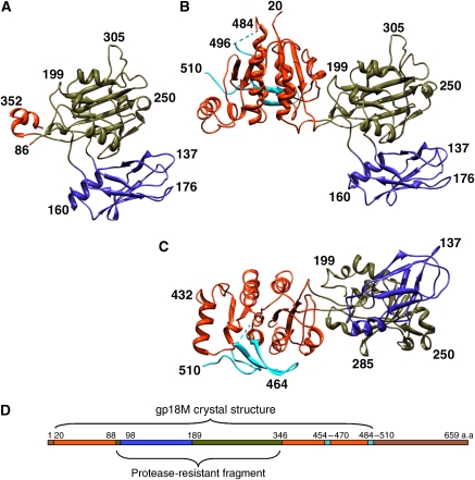
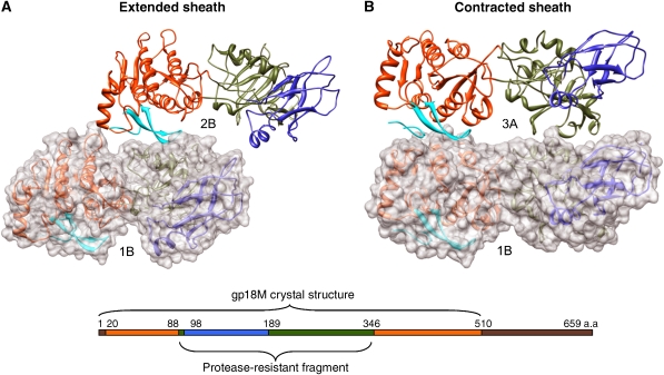
The contractile tail of bacteriophage T4 is a molecular machine that facilitates very high viral infection efficiency. Its major component is a tail sheath, which contracts during infection to less than half of its initial length. The sheath consists of 138 copies of the tail sheath protein, gene product (gp) 18, which surrounds the central non-contractile tail tube. The contraction of the sheath drives the tail tube through the outer membrane, creating a channel for the viral genome delivery. A crystal structure of about three quarters of gp18 has been determined and was fitted into cryo-electron microscopy reconstructions of the tail sheath before and after contraction. It was shown that during contraction, gp18 subunits slide over each other with no apparent change in their structure.


Figures

Figure 1
Figure 1
Structures of the gp18 deletion mutants. (A) A ribbon diagram of the protease resistant fragment (gp18PR). (B, C) A ribbon diagram of the gp18M mutant (¾ of the total protein length) in two orientations. The three domains are shown in blue (domain I), olive green (domain II) and orange red (domain III); the β-hairpin (residues 454–470) and the last 27 C-terminal residues of gp18M are shown in cyan. (D) Domain positions on the amino-acid sequence, using the same colour scheme as in (A), (B) and (C). Brown indicates the part of gp18 for which the atomic structure remains unknown.
Figure 2
Figure 2
Fit of the gp18M structure into (A) the extended (left) and (C) the contracted (right) tail cryo-EM densities. Shown for each conformation is a one-disk-thick slab (top left and right), a two-disk-thick slab (middle left and right), a closer view of the fit (bottom left and right) and the whole tail (center left and right). In both (A and C) top panels, the one-disk thick slab of density is shown with six gp18M molecules fitted into the density with their domains I, II and III coloured blue, olive green and orange red, respectively. In the two-disk-thick slabs of density shown in (A) and (C), the tail tube density is colored red and two sequential disks of the tail sheath are colored blue and green. In the closer view of the fit, four disks of the tail sheath are shown, with disks one and three colored green and disks two and four colored blue. In (B), the arrangement of the gp18M domains is shown in the linear sequence (top), in the ribbon diagram of the crystal structure (left) and in the structure fitted into the piece of density map that corresponds to full-length gp18 (right).
Figure 3
Figure 3
Relative position of two gp18 molecules belonging to the same helical strand in the extended and contracted tail sheath. (A) Four successive rings of electron density of the extended tail sheath with the densities of two gp18 molecules. (B) Superposition of the gp18 density extracted from the extended sheath onto the contracted sheath. The top panels in both (A) and (B) show the surface of the sheath, whereas the lower panel is a cut-away view showing the arrangement of the inner domains (brown). The cut-away plane is shown on a diagrammatic top view of the sheath with the direction of view indicated by an arrow. The gp18 molecule densities are colored as shown in the linear sequence diagram, with domains I, II, III and IV colored blue, green, orange and brown, respectively.
Figure 4
Figure 4
Connectivity between subunits in the extended and contracted tail sheath. The subunits that form three of the six neighbouring helices (pink, A; blue, B; and green, C) within the sheath are shown as surface representations of (A) the extended and (B) the contracted sheath. The successive hexameric disks are numbered 1, 2, 3, 4 and 5, with the disk being closest to the baseplate numbered 1. On the left is shown a surface representation side view of the tail. Immediately next to it is shown the whole view with a closer view of the three neighbouring helices (in pink, blue and green). In the same column as the closer view of the helical arrangement is shown, the schematic diagram of the arrangement of the three outer domains using the same colour scheme with each gp18 molecule is being represented by a cross. Right next to this is shown the arrangement of the inner helices consisting only of domains IV. This domain retains the connectivity between neighbouring subunits within each helix in both (A) the extended and (B) the contracted sheath. At the right of each panel is shown the top view of one disk of the tail sheath in the (A) extended and (B) contracted conformations. The three outer domains that correspond to the atomic structure are shown in ribbon representation. Domain IV, whose structure has not yet been determined, is shown as density segmented from the cryo-EM map.
Figure 5
Figure 5
Analysis of polymerization properties of gp18 deletion mutants. The mutants that can polymerize (including the full-length protein) are shown in blue and those that do not polymerize are shown in red. The gp18M construct is indicated by an arrow. The region of the sequence that influences polymerization properties is shown in the red rectangle and corresponds to the C-terminal 27 residues of the gp18M structure. Internal deletions are shown as thin lines, with deleted residues being indicated on top. The C-terminal mutations are shown with the three-letter amino-acid code.
Figure 6
Figure 6
Interactions between the gp18 subunits that involve β-hairpin (residues 454–470). Neighbouring gp18 subunits are shown for (A) extended and (B) contracted tail sheaths. One of the subunits is shown in transparent surface representation and both subunits are shown in ribbon representation. Gp18 domains colored as in linear sequence diagram with β-hairpin as well as the last 27-residue loop are shown in cyan. The interacting subunits correspond to subunits 1B and 2B of the extended tail sheath and subunits 1B and 3A for the contracted sheath according to the labelling in Figure 4.

Comment in

  • How viruses infect bacteria?
    Orlova EV. Orlova EV. EMBO J. 2009 Apr 8;28(7):797-8. doi: 10.1038/emboj.2009.71. EMBO J. 2009. PMID: 19352408 Free PMC article. No abstract available.

Similar articles

Cited by

References

    1. Ackermann H-W (2006) Classification of bacteriophages. In The Bacteriophages, Calendar R (ed), 2nd edn, pp 8–16. Oxford University Press: New York, NY
    1. Afonine PV, Grosse-Kunstleve RW, Adams PD (2005) The Phenix refinement framework. CCP4 Newsletter. Number 42, Contribution 8
    1. Arisaka F, Engel J, Horst K (1981) Contraction and dissociation of the bacteriophage T4 tail sheath induced by heat and urea. Prog Clin Biol Res 64: 365–379 - PubMed
    1. Arisaka F, Takeda S, Funane K, Nishijima N, Ishii S (1990) Structural studies of the contractile tail sheath protein of bacteriophage T4. 2. Structural analyses of the tail sheath protein, gp18, by limited proteolysis, immunoblotting, and immunoelectron microscopy. Biochemistry 29: 5057–5062 - PubMed
    1. Arisaka F, Tschopp J, van Driel R, Engel J (1979) Reassembly of the bacteriophage T4 tail from the core-baseplate and the monomeric sheath protein P18: a co-operative association process. J Mol Biol 132: 369–386 - PubMed

Publication types

MeSH terms

Substances