. 2009 Feb;5(2):e1000310.
doi: 10.1371/journal.ppat.1000310. Epub 2009 Feb 20.

Receptor complementation and mutagenesis reveal SR-BI as an essential HCV entry factor and functionally imply its intra- and extra-cellular domains

Affiliations

Receptor complementation and mutagenesis reveal SR-BI as an essential HCV entry factor and functionally imply its intra- and extra-cellular domains

Marlène Dreux et al. PLoS Pathog. 2009 Feb.

Abstract

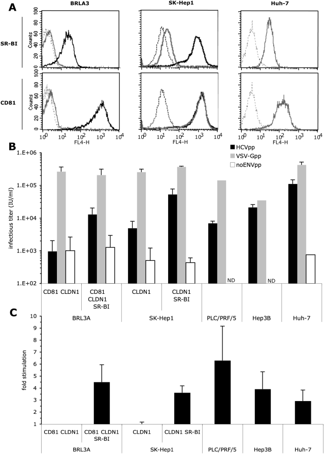
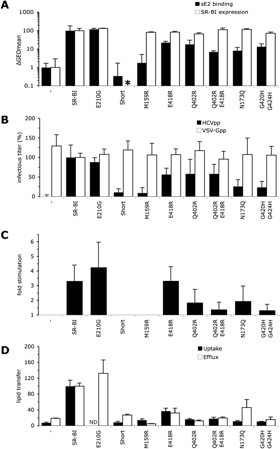
HCV entry into cells is a multi-step and slow process. It is believed that the initial capture of HCV particles by glycosaminoglycans and/or lipoprotein receptors is followed by coordinated interactions with the scavenger receptor class B type I (SR-BI), a major receptor of high-density lipoprotein (HDL), the CD81 tetraspanin, and the tight junction protein Claudin-1, ultimately leading to uptake and cellular penetration of HCV via low-pH endosomes. Several reports have indicated that HDL promotes HCV entry through interaction with SR-BI. This pathway remains largely elusive, although it was shown that HDL neither associates with HCV particles nor modulates HCV binding to SR-BI. In contrast to CD81 and Claudin-1, the importance of SR-BI has only been addressed indirectly because of lack of cells in which functional complementation assays with mutant receptors could be performed. Here we identified for the first time two cell types that supported HCVpp and HCVcc entry upon ectopic SR-BI expression. Remarkably, the undetectable expression of SR-BI in rat hepatoma cells allowed unambiguous investigation of human SR-BI functions during HCV entry. By expressing different SR-BI mutants in either cell line, our results revealed features of SR-BI intracellular domains that influence HCV infectivity without affecting receptor binding and stimulation of HCV entry induced by HDL/SR-BI interaction. Conversely, we identified positions of SR-BI ectodomain that, by altering HCV binding, inhibit entry. Finally, we characterized alternative ectodomain determinants that, by reducing SR-BI cholesterol uptake and efflux functions, abolish HDL-mediated infection-enhancement. Altogether, we demonstrate that SR-BI is an essential HCV entry factor. Moreover, our results highlight specific SR-BI determinants required during HCV entry and physiological lipid transfer functions hijacked by HCV to favor infection.


Conflict of interest statement

The authors have declared that no competing interests exist.

Figures

Figure 1
Figure 1. HCVpp entry upon ectopic hSR-BI co-expression with other HCV receptors.
(A) Endogenous and/or ectopic expression of hSR-BI and hCD81 in BRL3A, SK-Hep1, and Huh-7 cells was determined by flow cytometry. Parental cells were stained with hSR-BI (CLA-1 mAb, upper panel) or hCD81 (JS81 mAb, lower panel) antibodies (gray lines). The background of fluorescence was provided by staining the cells with the secondary antibodies only (dotted lines). Ectopic expression of hSR-BI in BRL3A and SK-Hep1 cells or of hCD81 in BRL3A cells was determined using the same antibodies (black lines). The results are representative of three independent experiments. (B) Results of HCV entry assays on BRL3A and SK-Hep1 cells ectopically expressing the indicated HCV receptors, and on PLC/PRF/5, Hep3B, and Huh-7 human hepatoma cells, endogenously expressing CD81, CLDN1, and SR-BI, using HCV pseudo-particles harboring H77-E1E2 glycoproteins (HCVpp), control viral particles harboring the VSV-G glycoprotein (VSV-Gpp; diluted 1/100), or no glycoprotein (noENVpp). The viral particles were produced in cell culture media devoid of serum lipoproteins. Results display average infectious titers, expressed as GFP IU/ml (mean±SD; n = 3). ND, not determined. (C) HCV entry assays using HCVpp produced in serum-free medium in the presence of 6 µg/ml cholesterol-HDL. The results show the fold increases of infection (mean±SD; n = 3) determined by calculating the ratios between average infectious titers determined in the presence or absence of HDL. No changes of infectivity with VSV-Gpp control particles were detected under these experimental conditions (data no shown), as reported previously .
Figure 2
Figure 2. HCVcc entry upon ectopic hSR-BI co-expression with other HCV receptors.
(A) Results of HCVcc entry assays, assessed by measuring intracellular HCV RNA level at 4, 12, and 72 hr post-infection, in SK-Hep1 cells ectopically expressing CLDN1 vs. CLDN1 and SR-BI, using cell culture–produced HCVcc in the absence or in the presence of NS3 protease inhibitor and NS5B-dependent RNA synthesis inhibitor (BILN2051 and 2′-C-methyl-adenosine, respectively, kindly provided by FV Chisari). Results are standardized with respect to the HCV RNA level obtained at 72 hr on non-treated SK-Hep1-CLDN1-SR-BI cells (mean±SD; n = 3). Intracellular HCV RNA levels of SK-Hep1-CLDN1-SR-BI was on average 440-fold lower than HCV RNA levels measured in Huh7.5 cell (4.7×10³±2.4×10² genome copies per µg cellular mRNA) using the same HCVcc supernatants. (B) Results of HCVcc entry assays, assessed at 72 hr post-infection, in SK-Hep1 cells ectopically expressing the indicated HCV entry factors, using cell culture–produced HCVcc in the absence (white bars) or presence (black bars) of 0.6 µg/ml cholesterol-HDL. Results are standardized with respect to HCV RNA level obtained in the absence of HDL on wt SR-BI–expressing SK-Hep1-CLDN1 cells (mean±SD; n = 5). Similar experiments in BRL3A-CD81-CLDN1-SR-BI cells allowed detection of HCV RNA at 72 hr, which was specifically increased upon treatment with HDL, and revealed differences upon expression of HCV entry factors (data not shown) consistent with the results obtained with HCVpp. However, kinetic experiments and use of replication inhibitors did not allow firm demonstration of HCV replication in these cells. (C) Dose-response curves for the SR-BI–dependent free cholesterol efflux and for HDL-CE uptake determined in BRL3A-CD81-CLDN1 and SKHep1-CLDN1 cells ectopically expressing, or not, hSR-BI, or in Fu5AH cells expressing high endogenous levels of rat SR-BI. Cholesterol efflux is expressed as the percentage of total labelled 3H-cholesterol released to the medium. Selective HDL-CE uptake is expressed as the percentage of labelled HDL-CE delivered to cells per µg of cell protein. The values represent the means ±SD of three experiments.
Figure 3
Figure 3. HCVpp entry in BRL3A-CD81-CLDN1 cells expressing SR-BI intracellular domain mutants.
(A) Cell surface expression (white bars) and sE2 binding (black bars) of SR-BI mutants/isoforms was determined using anti-SR-BI antibody (CLA-1 mAb) and soluble E2 protein (sE2), respectively. The results of cell surface expression, analyzed by flow cytometry of BRL3A-CD81-CLDN1 cells transduced with retroviral vectors carrying the indicated SR-BI mutants, are expressed as the average percentages of GEOmean (geometric mean) fluorescence shifts (mean±SD; n = 3) detected between mutant receptor–expressing cells and parental (—) cells, relative to cells expressing wild-type SR-BI (ca. 20-fold of GEOmean shift, see Figure 1A) set to 100. The results of sE2 binding are expressed as the average percentages of GEOmean fluorescence shifts (mean±SD; n = 3) detected in parental BRL3A cells (—) or in BRL3A cells only expressing the indicated SR-BI mutants that were incubated with sE2-containing medium vs. sE2-free medium, relative to cells expressing wild-type SR-BI (ca. 10-fold GEOmean shift, see Figure 6) set to 100. Cell surface expression of SR-BI mutants in these latter cells was similar to that detected in the SR-BI–expressing BRL3A-CD81-CLDN1 cells (data not shown). Cell surface expression of CD36 (*, data not shown) was verified using a CD36 antibody (FA6-152, abcam). (B) Effect of SR-BI mutations on infectivity of HCVpp produced in serum-free media. The results of infectivity (mean±SD; n = 5) are expressed relative to the infectious titers of HCVpp or of control VSV-Gpp particles determined on wt SR-BI–expressing BRL3A-CD81-CLDN1 cells (input ca. 104 GFP IU) set to 100. (C) Results of HCVpp infection-enhancement induced by HDL (6 µg/ml cholesterol-HDL), expressed as ratios between average infectious titers determined in the presence or absence of HDL (mean±SD; n = 5). No changes of infectivity of VSV-Gpp control particles were detected under these experimental conditions (data not shown), as reported previously . (D) Relative capacities of SR-BI mutants to mediate HDL-CE uptake (black bars) and free cholesterol efflux (white bars) relative to wt SR-BI set to 100 (mean±SD; n = 3). ND, not determined.
Figure 4
Figure 4. HCVcc entry in SK-Hep1-CLDN1 cells expressing SR-BI mutants.
The SR-BI mutants tested were the SR-BI/CD36 chimera and the SES mutants, altering SR-BI intracellular domain, the Q402R/E418R, N173Q, and G420H-G424H mutants, altering SR-BI ectodomain. (A) Effect of SR-BI mutations on infectivity of HCVcc produced under standard conditions and assessed by measuring intracellular HCV RNA level at 72 hr post-infection (see Figure 2A). The results of infectivity (mean±SD; n = 3) are expressed relative to HCVcc infection of wt SR-BI–expressing SK-Hep1-CLDN1 cells, which was set to 100. (B) Results of HCVcc infection-enhancement induced by HDL (0.6 µg/ml cholesterol-HDL) on wt SR-BI–expressing SK-Hep1-CLDN1 cells, expressed as ratios relative to infection in the absence of HDL of the same cells set to 100 (mean±SD; n = 3). Similar experiments in BRL3A-CD81-CLDN1 cells expressing these SR-BI mutants were performed (data not shown) and were consistent with the results obtained in SK-Hep1-CLDN1 cells.
Figure 5
Figure 5. HCVpp entry in BRL3A-CD81-CLDN1 cells expressing SR-BI ectodomain mutants.
(A) Cell surface expression (white bars) and sE2 binding (black bars) of SR-BI mutants/isoforms as determined using anti-SR-BI antibody (CLA-1 mAb) and soluble E2 protein (sE2), respectively. The results of cell surface expression, analyzed by flow cytometry of BRL3A-CD81-CLDN1 living cells transduced with retroviral vectors carrying the indicated SR-BI mutants, are expressed as the average percentages of GEOmean (geometric mean) fluorescence shifts (mean±SD; n = 3) detected between mutant receptor–expressing cells and parental (—) cells, relative to cells expressing wild-type SR-BI (ca. 20-fold of GEOmean shift, see Figure 1A) set to 100. The results of sE2 binding are expressed as the average percentages of GEOmean fluorescence shifts (mean±SD; n = 3) detected in parental BRL3A cells (—) or in BRL3A cells only expressing the indicated SR-BI mutants that were incubated with sE2-containing medium vs. sE2-free medium, relative to cells expressing wild-type SR-BI (ca. 10-fold of GEOmean shift, see Figure 6) set to 100. Cell surface expression of SR-BI mutants in these latter cells was similar to that detected in the SR-BI-expressing BRL3A-CD81-CLDN1 cells (data not shown). Cell surface expression of SR-BI-Short (*, data not shown) was verified by immuno-blotting using an antibody against SR-BI C-terminus (400-104, Novus) on surface-biotinylated proteins that were purified with streptavidin-coated beads. (B) Effect of SR-BI mutations on infectivity of HCVpp produced in serum-free media. The results of infectivity (mean±SD; n = 5) are expressed relative to the infectious titers of HCVpp or of control VSV-Gpp particles determined on wt SR-BI–expressing BRL3A-CD81-CLDN1 cells (input ca. 104 GFP i.u.), set to 100. (C) Results of HCVpp infection-enhancement induced by HDL (6 µg/ml cholesterol-HDL), expressed as ratios between average infectious titers determined in the presence or absence of HDL (mean±SD; n = 5). No changes of infectivity of VSV-Gpp control particles were detected under these experimental conditions (data not shown), as reported previously . (D) Relative capacities of SR-BI mutants to mediate HDL-CE uptake (black bars) and free cholesterol efflux (white bars) relative to wt SR-BI set to 100 (mean±SD; n = 3). ND, not determined.
Figure 6
Figure 6. Inhibition of sE2 binding to SR-BI by BLT-4 compound.
BRL3A cells expressing the indicated entry factors were incubated for 1 hr at 37°C with soluble E2 protein (sE2) (plain lines) or without sE2 (dotted lines) at saturating concentrations, after pre-incubation for 45 min at 37°C with (gray lines) or without (black lines) 50 µM BLT-4 (Chembridge), a SR-BI inhibitor , and with (left panels) or without (right panels) HDL (6 µg/ml cholesterol-HDL). SR-BI expression was not modified by the incubation with BLT-4 (data not shown). The data are representative of three independent experiments.

References

    1. Shepard CW, Finelli L, Alter MJ. Global epidemiology of hepatitis C virus infection. Lancet Infect Dis. 2005;5:558–567. - PubMed
    1. Bartosch B, Cosset F-L. Cell entry of hepatitis C virus. Virology. 2006;348:1–12. - PubMed
    1. Dubuisson J, Helle F, Cocquerel L. Early steps of the hepatitis C virus life cycle. Cell Microbiol. 2008;10:821–827. - PubMed
    1. von Hahn T, Rice CM. Hepatitis C virus entry. J Biol Chem. 2008;283:3689–3693. - PubMed
    1. Thomssen R, Bonk S, Propfe C, Heermann KH, Kochel HG, et al. Association of hepatitis C virus in human sera with beta-lipoprotein. Med Microbiol Immunol. 1992;181:293–300. - PubMed

Publication types

MeSH terms