. 2009 Feb;5(2):e1000387.
doi: 10.1371/journal.pgen.1000387. Epub 2009 Feb 20.

Poly (ADP-ribose) polymerase 1 is required for protein localization to Cajal body

Affiliations

Poly (ADP-ribose) polymerase 1 is required for protein localization to Cajal body

Elena Kotova et al. PLoS Genet. 2009 Feb.

Abstract

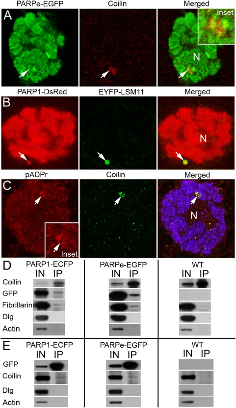
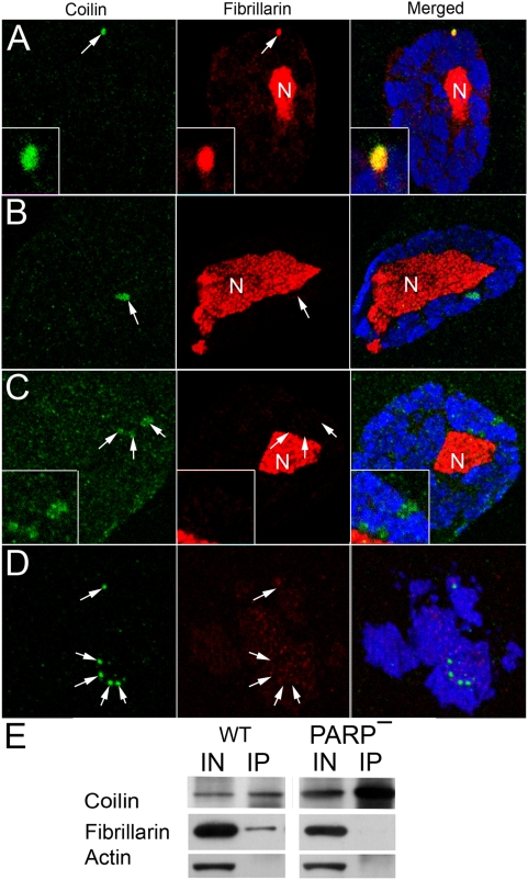
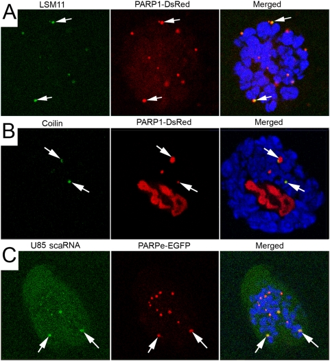
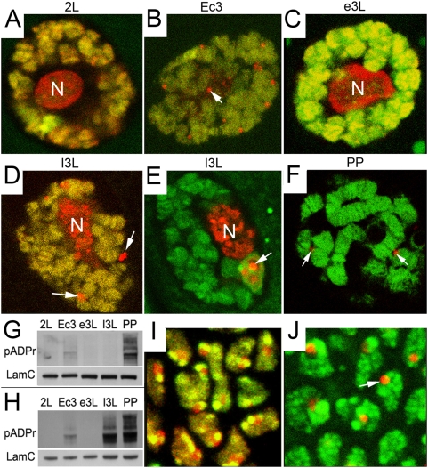
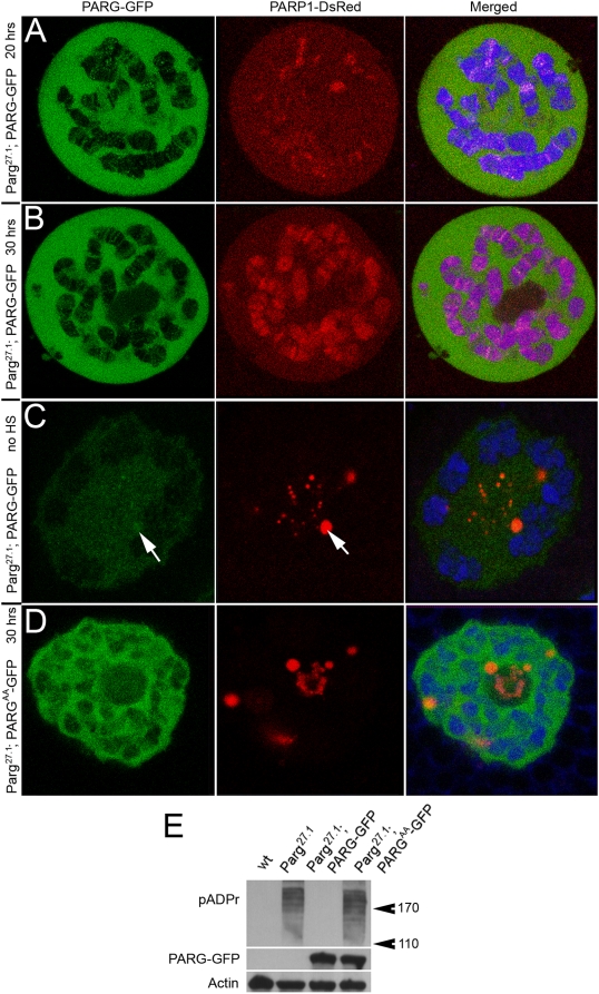
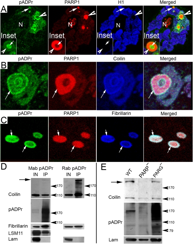
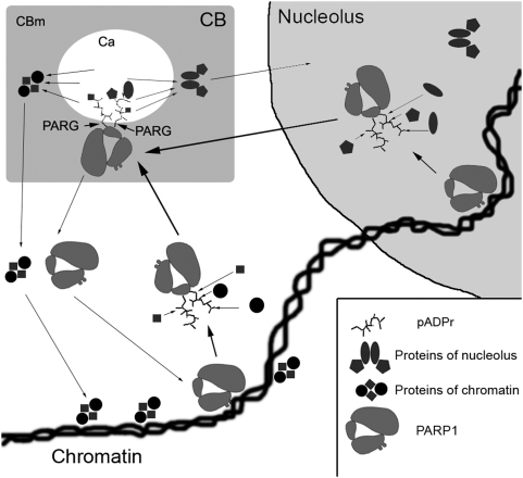
Recently, the nuclear protein known as Poly (ADP-ribose) Polymerase1 (PARP1) was shown to play a key role in regulating transcription of a number of genes and controlling the nuclear sub-organelle nucleolus. PARP1 enzyme is known to catalyze the transfer of ADP-ribose to a variety of nuclear proteins. At present, however, while we do know that the main acceptor for pADPr in vivo is PARP1 protein itself, by PARP1 automodification, the significance of PARP1 automodification for in vivo processes is not clear. Therefore, we investigated the roles of PARP1 auto ADP-ribosylation in dynamic nuclear processes during development. Specifically, we discovered that PARP1 automodification is required for shuttling key proteins into Cajal body (CB) by protein non-covalent interaction with pADPr in vivo. We hypothesize that PARP1 protein shuttling follows a chain of events whereby, first, most unmodified PARP1 protein molecules bind to chromatin and accumulate in nucleoli, but then, second, upon automodification with poly(ADP-ribose), PARP1 interacts non-covalently with a number of nuclear proteins such that the resulting protein-pADPr complex dissociates from chromatin into CB.


Conflict of interest statement

The authors have declared that no competing interests exist.

Figures

Figure 1
Figure 1. PARP protein is localized to Cajal body (CB) and histone locus body (HLB) and interacts with HLB/CB components.
N indicates nucleolus. A. The dissected salivary glands expressing PARPe-EGFP (green) transgenic construct were sectioned to ultra thin sections and stained with rabbit anti-Coilin antibody (red). Position of HLB/CB is indicated with arrow. Inset. Magnification of the chromatin block containing CB is shown. B. The dissected salivary glands co-expressing PARP1-DsRed (red) and EYFP-LSM11 (green) transgenic constructs were stained with the DNA binding dye Draq5 (blue). Position of HLB/CB is indicated with arrow. C. The dissected salivary glands from wild-type larvae were fixed and sectioned, followed by immunostaining with rabbit anti-pADPr (Red); Guinea Pig anti-Coilin antibody (green) and mouse anti-histone H1 (blue). Position of CB/HLB is indicated with arrow. Accumulation of pADPr in CB is clearly shown. Inset. Magnification of the chromatin block containing CB is shown. D. Immunoprecipitation assays using Guinea Pig anti-Coilin antibody. Drosophila stocks expressing PARP1-ECFP and PARPe-EGFP were used. Wild type Drosophila stock was used as a control. To detect protein on Western blots, the following antibodies were used: rabbit anti-Coilin; rabbit anti-GFP (to detect PARP1-ECFP and PARPe-EGFP); rabbit anti-Fibrillarin; mouse anti-Dlg; and mouse anti-Actin. E. Immunoprecipitation assays using rabbit anti-GFP antibody. Drosophila stock expressing PARP1-ECFP and PARPe-EGFP were used. Wild type Drosophila stock was used as a control. To detect protein on Western blots, the following antibodies were used: Guinea Pig anti-Coilin; mouse anti-GFP (to detect PARP1-ECFP and PARPe-EGFP); mouse anti-Fibrillarin; mouse anti-Dlg; and mouse anti-Actin.
Figure 2
Figure 2. PARP1 protein controls Cajal body integrity.
The following antibodies were used for immunostainings: Guinea Pig anti-Coilin (green); rabbit anti-Fibrillarin (red); and the DNA binding dye Draq5 (blue). N – nucleolus. The position of CB is indicated with arrow. Wild-type (A) and PARP1 mutant (B, C, D) salivary glands were used for immunostaining. E. Immunoprecipitation assays using mouse anti-Coilin antibody. The total protein extracts from wild-type (WT) and PARP1 mutant (PARP) flies were used. To detect protein on Western blots, rabbit anti-Coilin, rabbit anti-Fibrillarin, and mouse anti-Actin antibodies were used.
Figure 3
Figure 3. The inhibition of PARG function induces ectopic CBs and relocation of PARP1 protein from chromatin into CBs.
(A, B) The salivary glands dissected from the Parg27.1 mutants expressing PARP1-DsRed (red) transgene. DNA was stained with Draq5 dye. Antibody against LSM11 protein (green) (A) and anti-Coilin (green) (B) were used to detect Cajal bodies. N – nucleolus. (C) Fluorescent in situ hybridization of U85 scaRNA. The salivary glands dissected from the Parg27.1 mutants expressing PARPe-EGFP transgene. U85 scaRNA probe (green) detects CBLP. Rabbit antibody against GFP protein (red) detects PARPe-EGFP. To visualize chromatin (blue), mouse anti-histone H1 antibody was used. Arrows indicate CBLP with strong colocalization of U85 scaRNA and PARP protein.
Figure 4
Figure 4. Ecdysteroid hormone controls PARP1 protein localization and activation in vivo.
Localization of PARP1-DsRed (red) protein was analyzed by confocal microscopy in live Parg27.1 mutant Drosophila tissues. DNA was stained with Draq5 reagent (green). PARP-containing nucleoplasmic bodies are indicated with arrows. Salivary gland nuclei in late second-instar larvae (A), in ecdysis III (B), in early third-instar (C), late third-instar (D, E), and late prepupae (F) are shown. (G–H) Western blot analysis of the pADPr accumulation in wild- type (G) and Parg27.1 mutant animals (H) during development is shown. Mouse anti-pADPr antibody was used. Mouse antibody against Lamin C was used as a loading control. (I–J) The comparison of the PARP1-DsRed protein localization in diploid tissues (wing imaginal disk) of wild-type (I) and Parg27.1 mutant (J), late third-instar larvae, is shown.
Figure 5
Figure 5. The ectopic expression of recombinant PARG-EGFP protein in Parg27.1 mutant animals rescues PARP1-DsRed localization to chromatin.
The expression of UAS::PARG-EGFP transgene was induced in early second-instar larvae (A, B), or in early third-instar larvae (C), using hsp::Gal4 driver induction. Salivary glands were dissected and analyzed by confocal microscopy after 20 hrs following Gal4 induction (A, C) and after 30 hrs following Gal4 induction (B). PARG-EGFP protein is green; PARP1-DsRed is red, and DNA (Draq5) is blue. An arrow indicates colocalization of PARG-EGFP and PARP1-DsRed in the CB-like particle. D. The expression of recombinant catalytically inactive PARGAA-EGFP protein in Parg27.1 mutant animals does not rescue PARP1-DsRed protein localization to chromatin (after 30 hrs following Gal4 induction). E. Western blot hybridization was used to detect the accumulation of pADPr in Parg27.1 mutant and Parg27.1 mutant expressing PARGAA-EGFP protein, but not in wild-type and Parg27.1 mutant expressing PARG-EGFP protein.
Figure 6
Figure 6. The components of Cajal body are targets for pADPr.
(A) The immunostaining of thick sections prepared from Parg27.1 mutant salivary gland is shown. “Free” nucleoplasmic bodies (arrow) and “chromatin-embedded” CBs (arrowhead) are shown. Specific antibodies were used: rabbit anti-pADPr (green) and mouse anti-histone H1 (blue). PARP1-DsRed (red) was visualized by DsRed autofluorescence. Inset. The single CB-like particle is magnified. Arrow indicates accumulation of pADPr in the CB cavity. Arrowhead shows enrichment of PARP1 protein in the CB matrix. (B, C) Immunostaining of Parg27.1 mutant salivary gland is shown. Specific antibodies were used: mouse (10H) anti-pADPr (green) (B–C); Guinea Pig anti-Coilin (blue) (B) and rabbit anti-Fibrillarin (C). PARP1-DsRed (red) was visualized by DsRed autofluorescence. (B) The single chromatin-embedded CB-like particle is presented. Arrow indicates the colocalization of Coilin and pADPr on periphery of CB. (C) Three CBs are shown. Arrows show the colocalization of Fibrillarin and pADPr on periphery of CBs. (D) Immunoprecipitation assays using mouse and rabbit antibody against pADPr. Wild-type Drosophila stock was used to prepare protein extracts. To detect protein on Western blots, the following antibodies were used: Guinea Pig anti-Coilin, mouse anti-pADPr, rabbit anti-Fibrillarin, rabbit anti-LSM11 and mouse anti-Lamin C. Arrow indicates hypermodified isoform of Coilin. (E) Western blot analysis of total protein extracts from wild-type (WT), Parp1 (PARP) and Parg (PARG) mutant flies was performed. Rabbit anti-Coilin antibody was used. Arrow indicates hypermodified isoform of Coilin.
Figure 7
Figure 7. A model of protein delivery to Cajal body by PARP1 shuttling is shown.
(1) PARP1 protein is localized in chromatin and nucleoli. (2) Upon activation, PARP1 automodifies and (3) gains the ability to bind by pADPr a number of proteins with pADPr-binding domain. (4) Whole complex consisting of automodified PARP1 and proteins seated on pADPr migrates into Cajal bodies. (5) In CB, complex is disassembled as a result of cleavage of pADPr and released proteins are recycled. PARP1, pADPr protein-complexes of chromatin and nucleolus are indicated. CBm = Cajal body matrix, and Ca = Cajal body cavity.

Similar articles

Cited by

References

    1. De Murcia G, Shall S. From DNA damage and stress signalling to cell death Poly (ADP-Ribosylation) Reactions. New York: Oxford University Press; 2000.
    1. D'Amours D, Desnoyers S, D'Silva I, Poirier GG. Poly(ADP-ribosyl)ation reactions in the regulation of nuclear functions. Biochem J. 1999;342:249–268. - PMC - PubMed
    1. Meder VS, Boeglin M, de Murcia G, Schreiber V. PARP-1 and PARP-2 interact with nucleophosmin/B23 and accumulate in transcriptionally active nucleoli. J Cell Sci. 2005;118:211–222. - PubMed
    1. Molinete M, Vermeulen W, Burkle A, Menissier-de Murcia J, Kupper JH, et al. Overproduction of the poly(ADP-ribose) polymerase DNA-binding domain blocks alkylation-induced DNA repair synthesis in mammalian cells. EMBO J. 1993;12:2109–2117. - PMC - PubMed
    1. Pinnola A, Naumova N, Shah M, Tulin AV. Nucleosomal core histones mediate dynamic regulation of PARP1 protein binding to chromatin and induction of PARP1 enzymatic activity. J Biol Chem. 2007;282:32511–32519. - PubMed

Publication types

MeSH terms

Substances