. 2009 Mar;15(3):89-100.
doi: 10.1016/j.molmed.2009.01.001. Epub 2009 Feb 21.

Calcium signaling and neurodegenerative diseases

Affiliations

Calcium signaling and neurodegenerative diseases

Ilya Bezprozvanny. Trends Mol Med. 2009 Mar.

Abstract

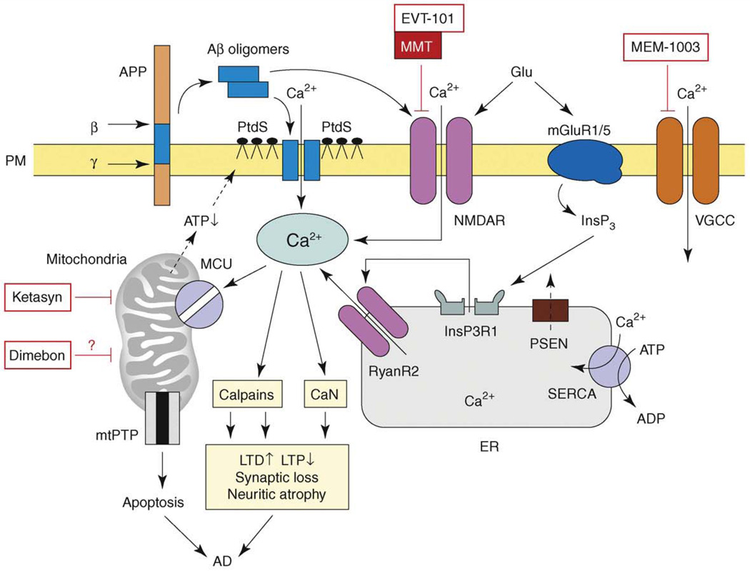
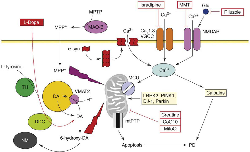
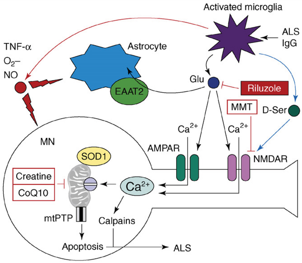
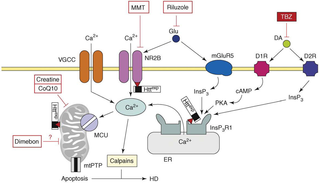
Neurodegenerative disorders, such as Alzheimer's disease (AD), Parkinson's disease (PD), amyotrophic lateral sclerosis (ALS), Huntington's disease (HD) and spinocerebellar ataxias (SCAs), present an enormous medical, social, financial and scientific problem. Recent evidence indicates that neuronal calcium (Ca2+) signaling is abnormal in many of these disorders. Similar, but less severe, changes in neuronal Ca2+ signaling occur as a result of the normal aging process. The role of aberrant neuronal Ca2+ signaling in the pathogenesis of neurodegenerative disorders is discussed here. The potential utility of Ca2+ blockers for treatment of these disorders is also highlighted. It is reasoned that Ca2+ blockers will be most beneficial clinically when used in combination with other disease-specific therapeutic approaches.


Conflict of interest statement

Disclosure statement

The author has no conflicts of interest to declare.

Figures

Figure 1
Figure 1
The model of Ca2+ dysregulation in AD. Sequential cleavages of β-amyloid precursor protein (APP) by β-secretase (β) and γ-secretase (γ) generate amyloid β-peptide (Aβ). Aβ forms oligomers, which can insert into the plasma membrane and form Ca2+-permeable pores. The association of Aβ oligomers with the plasma membrane is facilitated by binding to surface phosphatidylserine (PtdS); age and Ca2+-related mitochondrial impairment leads to ATP depletion and might trigger flipping of PtdS from the inner portion of the plasma membrane to the cell surface. Reduction in ATP levels and loss of membrane integrity causes membrane depolarization, which leads to facilitation of Ca2+ influx through NMDAR and VGCC. Aβ oligomers can also affect activity of NMDAR, AMPAR and VGCC directly. Glutamate stimulates activation of mGluR1/5 receptors, production of InsP3 and InsP3-mediated Ca2+ release from the ER. Presenilins (PS) function as an ER Ca2+-leak channels and many FAD mutations impair Ca2+-leak-channel function of PS, resulting in excessive accumulation of Ca2+ in the ER. Increased ER Ca2+ levels result in enhanced Ca2+ release through InsP3-gated InsP3R1 and Ca2+-gated RyanR2. PS might also modulate activity of InsP3R, RyanR and SERCA pump directly. Elevated cytosolic Ca2+ levels result in the activation of calcineurin (CaN) and calpains and lead to facilitation of LTD, inhibition of LTP, modification of neuronal cytoskeleton, synaptic loss and neuritic atrophy. Excessive Ca2+ is taken up by mitochondria through mitochondrial Ca2+ uniporter (MCU), eventually leading to opening of mitochondrial permeability-transition pore (mtPTP) and apoptosis. The NMDAR inhibitor memantine (MMT) is approved for the treatment of AD and the NR2B-specific antagonist EVT-101 was recently developed for AD treatment. ‘CNS-optimized’ L-type VGCC inhibitor MEM-1003, putative ‘mitochondrial agent’ Dimebon and ‘mitochondrial energizer’ Ketasyn are in clinical trials for AD. Adapted from [6].
Figure 2
Figure 2
The model of Ca2+ dysregulation in PD. Continuous Ca2+ influx to SNc neurons is mediated by CaV1.3 L-type voltage-gated Ca2+ channels (CaV1.3). In response to glutamate, Ca2+ influx is mediated by NMDA receptors (NMDAR). Alpha-synuclein forms aggregates (protofibrils) which may form Ca2+-permeable channels in the plasma membrane. Elevated cytosolic Ca2+ is transported into mitochondria through the activity of mitochondrial Ca2+ uniporter (MCU). Dopamine (DA) is generated from L-tyrosine by the action of tyrosine hydroxylase (TH) and is loaded into the synaptic vesicles by the activity of the DA/H+ cotransporter (VMAT2). Cytosolic DA is oxidized to 6-hydroxy-DA, which causes damage to proteins and mitochondria by oxidative stress. The products of DA oxidation accumulate as neuromelanin (NM). Cumulative damage to mitochondria resulting from Ca2+ overload and DA-mediated oxidative stress leads to an opening of mtPTP and apoptotic cell death of SNc neurons in PD. An importance of mitochondria is highlighted by several mitochondria-related genes (e.g. LRRK2, PINK1, DJ-1, Parkin), which are mutated in familial PD. In a chemical model of PD, the toxin 1-methyl-4-phenyl-1,2,3,6-tetrahydropyridine (MPTP) is converted to the 1-methyl-4-phenylpyridinium (MPP+) by the glial enzyme monoamine oxidase B (MAO-B). MPP+ enters SNc neurons and potently inhibits mitochondrial complex I, causing selective cell death of SNc neurons. The US FDA-approved treatment for PD is levodopa (L-dopa), which is converted to DA by aromatic L-amino acid decarboxylase (DCC) inside SNc neurons. Generated DA is loaded to synaptic vesicles and alleviates symptoms of PD temporarily. The drugs tested or in PD clinical trials currently are ‘mitochondrial stabilizers’ (creatine, CoQ10, MitoQ), NMDAR antagonist memantine (MMT), antiglutamate agent riluzole and L-type VGCC inhibitor isradipine.
Figure 3
Figure 3
The model of excitotoxicity and Ca2+ dysregulation in ALS. The pathogenic cascade in ALS involves interactions among activated microglia, astrocytes and motor neurons (MNs). In an experimental situation, microglia can be activated by antiserum collected from ALS patients (ALS IgG). Activated microglia release pro-inflammatory factors TNF-α, NO and O2. Activated microglia also release large amounts of glutamate, which causes activation of AMPA and NMDA receptors on MNs. Activated microglia also release D-serine, which further sensitizes NMDAR to glutamate activation. Astrocytes express glutamate-uptake transporter EAAT2, which is involved in clearing glutamate from the extracellular space. Ca2+ influx by Ca2+-permeable AMPA receptors and NMDA receptors results in mitochondrial Ca2+ overload, mitochondrial swelling, opening of mitochondrial permeability-transition pore (mtPTP) and apoptosis of MNs. Mutant SOD1 binds to MN mitochondria and further impairs their ability to handle Ca2+ load. Antiglutamate agent riluzole is approved by the US FDA for treatment of ALS. NMDAR antagonist memantine (MMT) and ‘mitochondrial stabilizers’ creatine and CoQ10 are in ALS clinical trials currently.
Figure 4
Figure 4
The model of Ca2+ dysregulation in HD. In HD MSN, the Httexp perturbs Ca2+ signaling through several synergistic mechanisms. Httexp enhances function of NR2B-containing NMDAR, probably by promoting trafficking to the plasma membrane. Httexp binds strongly to the InsP3R1C terminus and sensitizes the InsP3R1 to activation by InsP3. The low levels of glutamate released from corticostriatal projection neurons lead to supranormal Ca2+ influx by NMDAR and Ca2+ release through the InsP3R1. Additional Ca2+ influx to MSN is mediated by voltage-gated Ca2+ channels (VGCCs). Dopamine released from midbrain dopaminergic neurons stimulates D1-class and D2-class DARs, which are expressed abundantly in MSNs. D1-class DARs are coupled to activation of adenyl cyclase, increase in cAMP levels and activation of PKA. PKA potentiates glutamate-induced Ca2+ signals by facilitating the activity of NMDAR and InsP3R1. D2 receptors are coupled directly to InsP3 production and activation of InsP3R1. Supranormal Ca2+ signals activate calpain, which cleave Httexp and other substrates. Excessive cytosolic Ca2+ signals result in mitochondrial Ca2+ uptake by MCU, which eventually triggers mtPTP opening and apoptosis. The mitochondrial Ca2+ handling is further destabilized by direct association of Httexp with mitochondria. Antidopamine agent tetrabenazine (TBZ) is approved by the US FDA for symptomatic treatment of HD. NMDAR antagonist memantine (MMT), putative ‘mitochondrial agent’ Dimebon and ‘mitochondrial stabilizers’ creatine and CoQ10 are tested in HD clinical trials. Antiglutamate agent riluzole was tested and failed [66]. Adapted from [77].

References

    1. Toescu EC, Verkhratsky A. The importance of being subtle: small changes in calcium homeostasis control cognitive decline in normal aging. Aging Cell. 2007;6:267–273. - PubMed
    1. Gant JC, et al. Early and simultaneous emergence of multiple hippocampal biomarkers of aging is mediated by Ca2+-induced Ca2+ release. J. Neurosci. 2006;26:3482–3490. - PMC - PubMed
    1. Foster TC. Calcium homeostasis and modulation of synaptic plasticity in the aged brain. Aging Cell. 2007;6:319–325. - PubMed
    1. Hardy J, Selkoe DJ. The amyloid hypothesis of Alzheimer’s disease: progress and problems on the road to therapeutics. Science. 2002;297:353–356. - PubMed
    1. Seabrook GR, et al. Beyond amyloid: the next generation of Alzheimer’s disease therapeutics. Mol. Interv. 2007;7:261–270. - PubMed

Publication types