. 2009 Apr;136(4):1288-96.
doi: 10.1053/j.gastro.2008.12.037. Epub 2008 Dec 13.

Amphiregulin-deficient mice develop spasmolytic polypeptide expressing metaplasia and intestinal metaplasia

Affiliations

Amphiregulin-deficient mice develop spasmolytic polypeptide expressing metaplasia and intestinal metaplasia

Ki Taek Nam et al. Gastroenterology. 2009 Apr.

Abstract

Background & aims: The loss of parietal cells from the fundic mucosa leads to the emergence of metaplastic lineages associated with an increased susceptibility to neoplastic transformation. Both intestinal metaplasia (IM) and spasmolytic polypeptide (TFF2/SP) expressing metaplasia (SPEM) have been identified in human stomach, but only SPEM is present in most mouse models of gastric metaplasia. We previously determined that loss of amphiregulin (AR) promotes SPEM induced by acute oxyntic atrophy. We have now examined whether SPEM in the AR-/- mouse predisposes the stomach to gastric neoplasia.

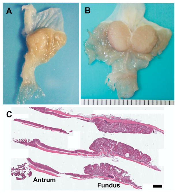
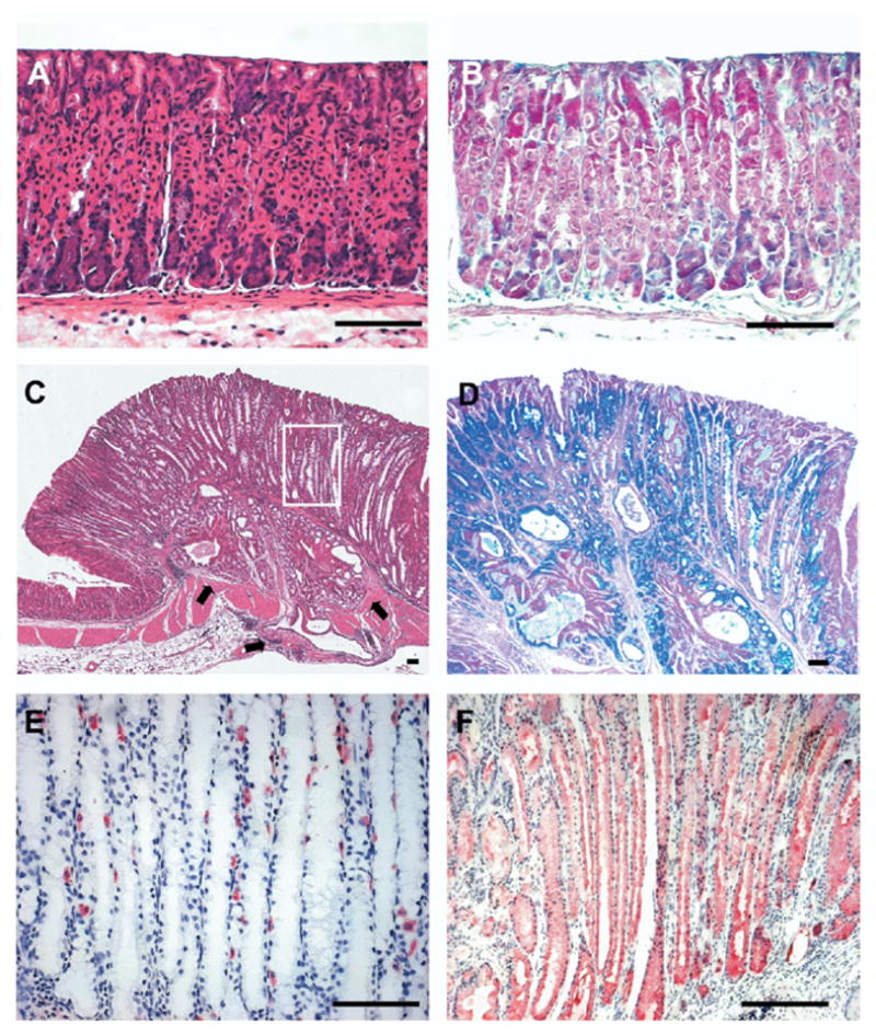
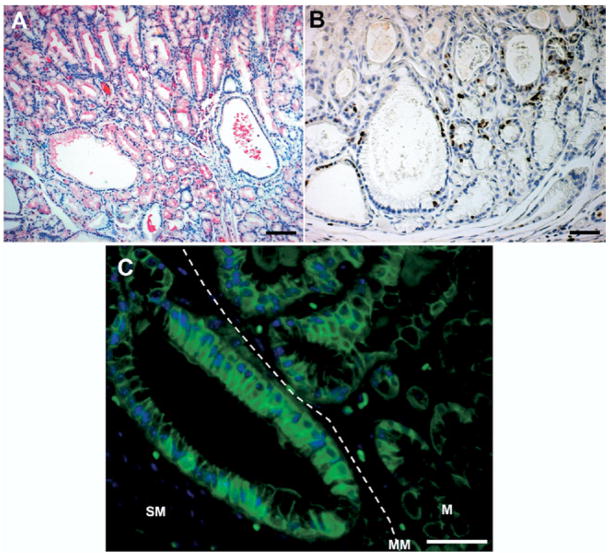
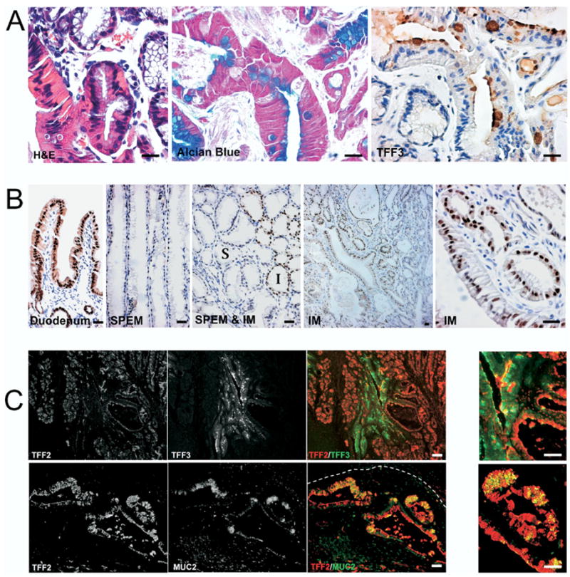
Methods: Gross pathology of 18-month-old wild-type, AR-/-, and TGF-alpha-/- mice were examined. Ki-67, beta-catenin, Pdx-1, TFF3, and TFF2/SP expression was analyzed by immunohistochemistry. Metaplastic gastric mucosa was analyzed by dual immunostaining for TFF2/SP with MUC2 or TFF3.

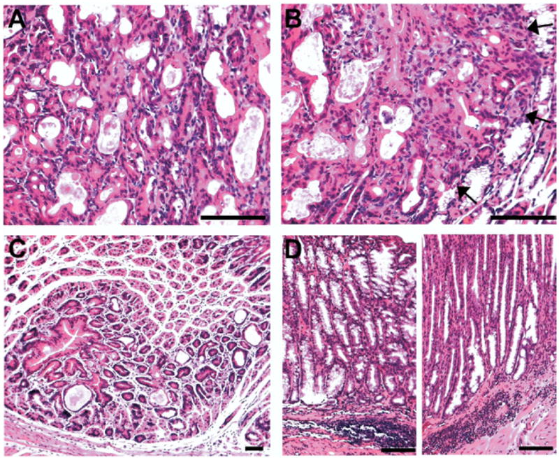
Results: By 18 months of age, more than 70% of AR-/- mice developed SPEM while 42% showed goblet cell IM labeled with MUC2, TFF3, and Pdx-1. A total of 28% had invasive gastric lesions in the fundus. No antral abnormalities were observed in AR-/- mice. Metaplastic cell lineages in AR-/- mice showed increases in cell proliferation and cytosolic beta-catenin expression. Dual staining for TFF2/SP with MUC2 or TFF3 showed glands containing both SPEM and IM with intervening cells expressing both TFF2/SP and MUC2 or TFF2/SP and TFF3.

Conclusions: AR-/- mice develop SPEM, which gives rise to goblet cell IM and invasive fundic dysplastic lesions. The AR-/- mouse represents the first mouse model for spontaneous development of fundic SPEM with progression to IM.


Conflict of interest statement

Conflicts of interest

The authors disclose no conflicts.

Figures

Figure 1
Figure 1
AR-deficient mice develop gross fundic tumors. Gross appearance of (A) a 10-month-old AR-deficient mouse stomach and (B) an 18-month-old AR-deficient mouse stomach. (A) The 10-month-old mice showed visible nodules arising in the fundic area, while (B) 18-month-old mice showed grossly visible tumors in the fundic area. (C) H&E staining of the stomach of an 18-month-old AR-deficient mouse. Note the vastly expanded fundic mucosa (Fundus) in contrast to the normal antral (Antrum) morphology in the histologic sections of the stomach. Bar = 1 mm.
Figure 2
Figure 2
Characterization of expanded fundic glands in 18-month-old AR-deficient mice. (A and B) Histologic sections of normal fundus stained with either (A) H&E or (B) periodic acid–Schiff/Alcian blue. (CF) Histologic sections of fundus from an 18-month-old AR-deficient mouse stained with (C) H&E, (D) periodic acid–Schiff/Alcian blue, (E) anti–H/K-adenosine triphosphatase, or (F) anti-TFF2. AR-deficient mice showed extensive replacement of the normal gland cell lineages by columnar mucous cells that were both periodic acid–Schiff and Alcian blue positive. Note the invasion of the metaplastic glands into muscle layer with cystically dilated glands (arrows in C). (E and F) Staining in serial sections of fundic gland region outlined in the white box in C showed severe loss of parietal cells (E) and replacement of virtually the entire mucosal gland length with intensely staining TFF2/SP-expressing cells. Bar = 100 μm.
Figure 3
Figure 3
Characteristics of invasive fundic glands in 18-month-old AR-deficient mice. To characterize the invasive regions of the metaplastic mucosa, sections were stained for (A) TFF2/SP, (B) Ki-67, and (C) β-catenin. (A) TFF2/SP-positive glands also were present in the invasive fundic gland area. (B) Note the Ki-67–positive cells in the invasive area, especially in cystic lesions. (C) Membrane localized β-catenin was detected in the glands within the mucosal area (M), but note the number of cells containing cytoplasmic β-catenin in submucosal (SM) invasive glands deep to the muscularis mucosa (MM). Bar = 50 μm.
Figure 4
Figure 4
Histopathologic analysis of representative fundic lesions in AR-deficient mice. Sections of fundic stomach from 18-month-old AR-deficient mice were stained with H&E and examined for histopathologic findings. (A and B) Invasive areas with histologic features of dysplasia. (B) Note the expanding dysplasia invading into the adjacent SPEM regions (arrows). (C) Fundic hyperplasia. Note the polypoid growth pattern of foveolar cells. Parietal cells around the polyp were normal. (D) Submucosal infiltration of inflammatory cells. Note the inflammatory cells were locally infiltrated in the submucosa only. There were no inflammatory infiltrates observed in the mucosa. Bar = 50 μm.
Figure 5
Figure 5
Serum levels of cytokines determined in AR- and TGF-α–deficient mice. Serum levels of IL-2, TNF-α, interferon gamma (IFN-γ), IL-5, and IL-4 were determined in 10-month-old WT, AR-deficient, and TGF-α–deficient mice. Both TGF-α and AR knockout mice showed significant elevations in all 5 cytokines compared with WT mice (*P < .01). In addition, levels of IL-5 were significantly elevated in AR−/− mice compared with TGF-α −/− (#P < .01).
Figure 6
Figure 6
Cytokine mRNA expression levels in gastric tissue from AR-and TGF-α–deficient mice. Total RNA was prepared from gastric tissue of 10-month-old WT, AR-deficient, and TGF-α–deficient mice. Cyto-kine mRNA expression was determined by quantitative reverse-transcription polymerase chain reaction for IL-2, IL-4, IL-5, TNF-α, and interferon gamma (INF-γ). No significant differences were observed between WT and TGF-α–deficient mice. However, significant elevations in IL-2, IL-4, IL-5, and TNF-α expression were observed in the gastric mucosa of AR-deficient mice compared with WT and TGF-α–deficient mice (*P < .05, **P < .01).
Figure 7
Figure 7
SPEM progresses to intestinal metaplasia in 18-month-old AR-deficient mice. (A) To identify goblet cell IM, sections of fundic stomach from 18 month-old AR-deficient mice were stained for H&E, Alcian blue, and TFF3 immunohistochemistry in adjacent sections. The stains show Alcian blue and TFF3-positive goblet cells within the mucosa. (B) To investigate the characteristics of goblet cell IM in stomach, sections from AR-deficient mice fundic mucosa were immunostained for the duodenal transcription factor Pdx-1. While Pdx-1 was not expressed in the SPEM regions, IM regions showed cells with prominent nuclear expression of Pdx-1. Note that no expression of Pdx-1 was observed in SPEM (S) in the surrounding regions of the IM (I). (C) To investigate IM development in the setting of SPEM, fundic sections from 18-month-old AR-deficient mice were dual stained for immunofluorescence labeling of TFF2/SP with TFF3 or TFF2/SP with MUC2. Note the TFF3-positive goblet cells appeared within TFF2/SP-positive glands (upper panels). In the invasive area (white dashed line indicates the position of the muscularis mucosa), cells staining for both TFF2/SP and MUC2 were observed in cystically dilated glands (lower panels). Panels at the far right show higher-magnification views of merged fluorescence images showing cells with dual labeling for either TFF2 with TFF3 or TFF2 with MUC2. Bar = 25 μm.

References

    1. Karam SM, Leblond CP. Dynamics of epithelial cells in the corpus of the mouse stomach. I. Identification of proliferative cell types and pinpointing of the stem cell. Anat Rec. 1993;236:259–279. - PubMed
    1. Karam SM, Leblond CP. Dynamics of epithelial cells in the corpus of the mouse stomach. III. Inward migration of neck cells followed by progressive transformation into zymogenic cells. Anat Rec. 1993;236:297–313. - PubMed
    1. El-Zimaity HM, Ota H, Graham DY, et al. Patterns of gastric atrophy in intestinal type gastric carcinoma. Cancer. 2002;94:1428–1436. - PubMed
    1. Correa P. A human model of gastric carcinogenesis. Cancer Res. 1988;48:3554–3560. - PubMed
    1. Filipe MI, Munoz N, Matko I, et al. Intestinal metaplasia types and the risk of gastric cancer: a cohort study in Slovenia. Int J Cancer. 1994;57:324–329. - PubMed

Publication types

MeSH terms