. 2009 Sep;5(3):305-15.
doi: 10.1016/j.nano.2008.11.003. Epub 2009 Feb 11.

Factorial analyses of photopolymerizable thermoresponsive composite hydrogels for protein delivery

Affiliations

Factorial analyses of photopolymerizable thermoresponsive composite hydrogels for protein delivery

Abhimanyu Sabnis et al. Nanomedicine. 2009 Sep.

Abstract

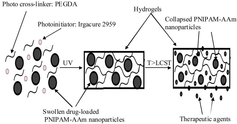
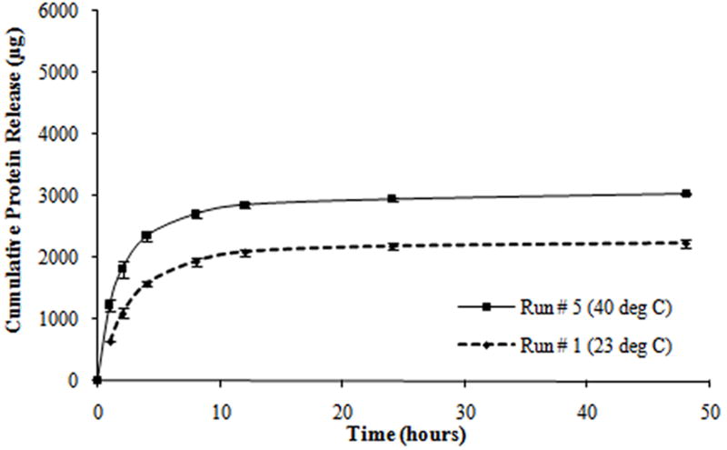
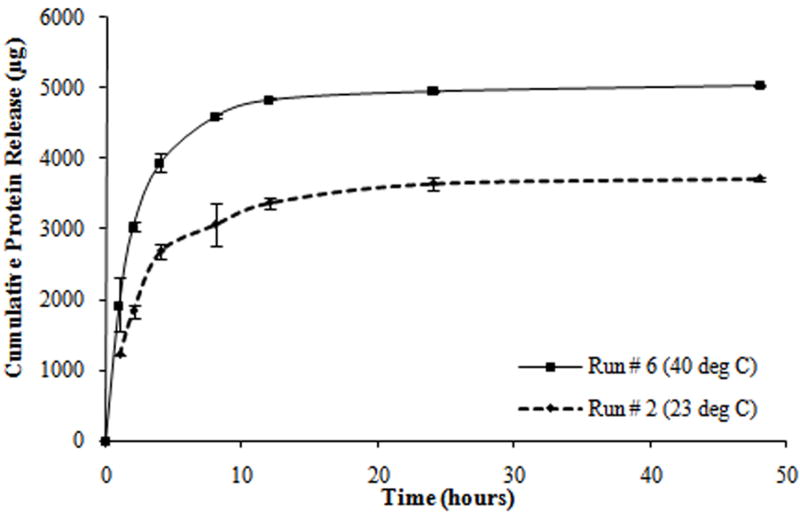
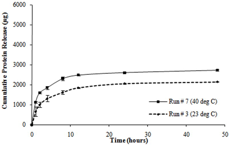
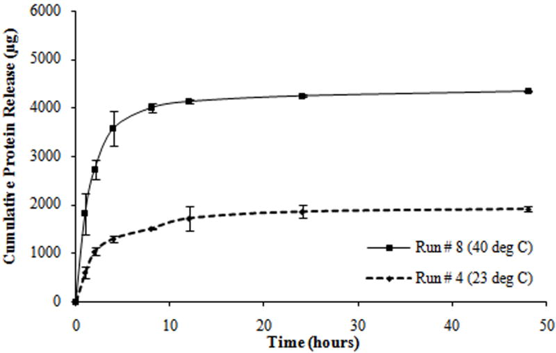
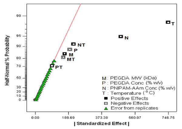
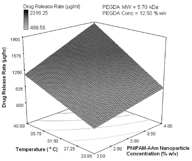
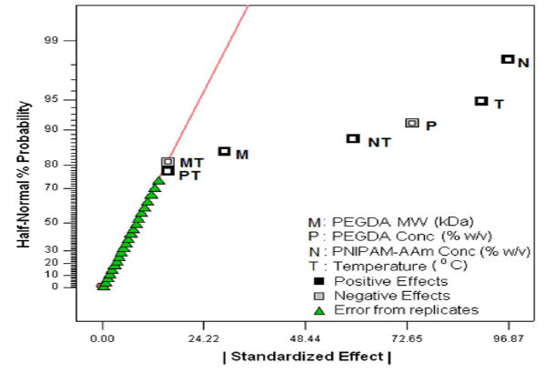
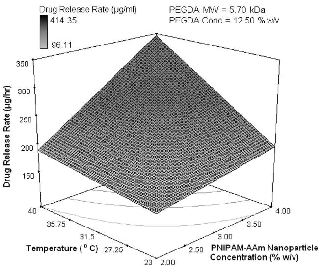
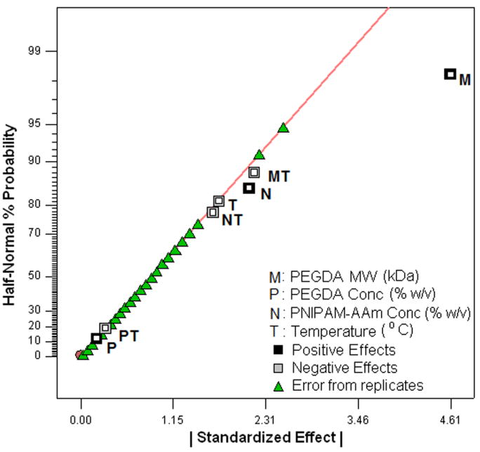
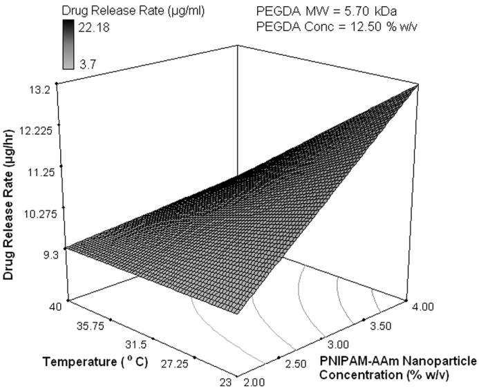
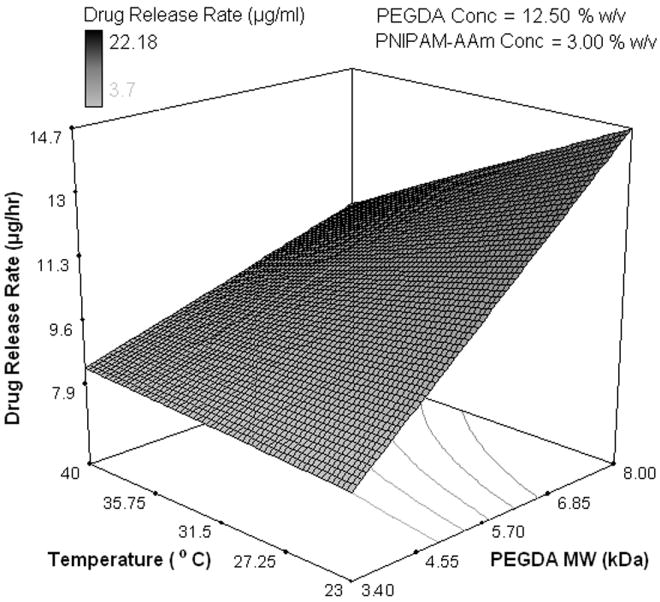
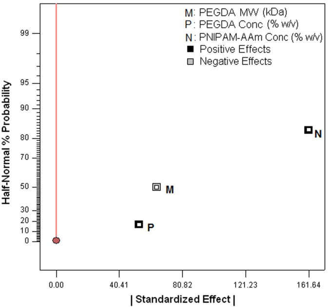
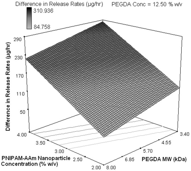
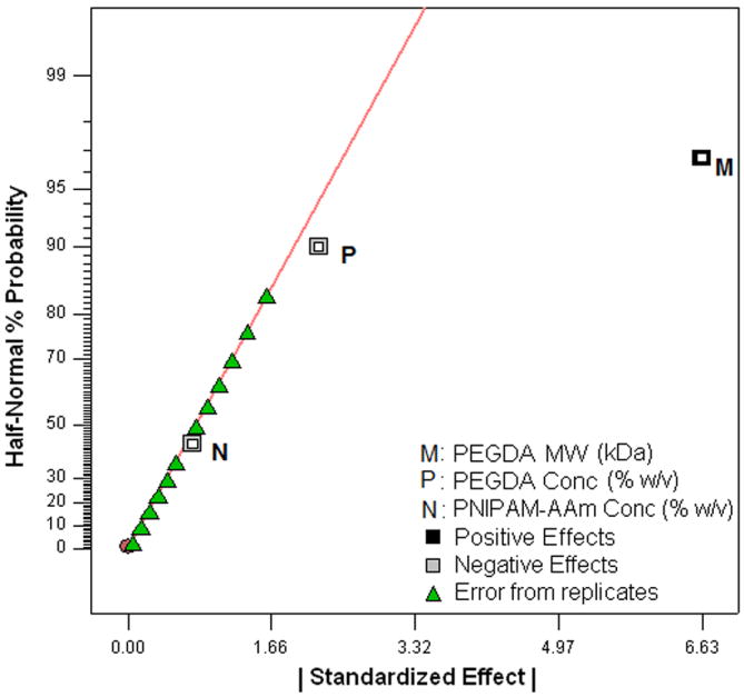
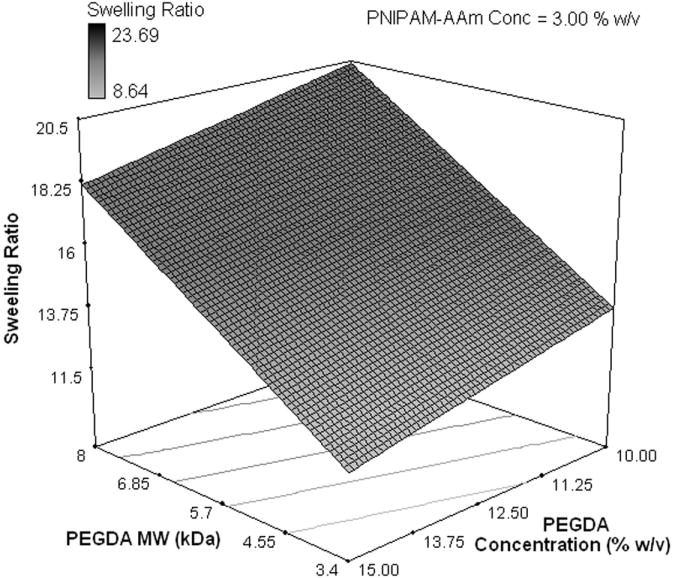
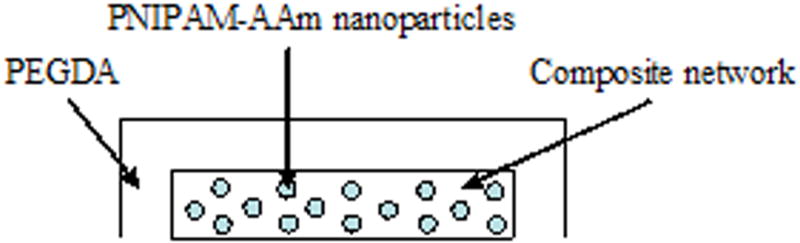
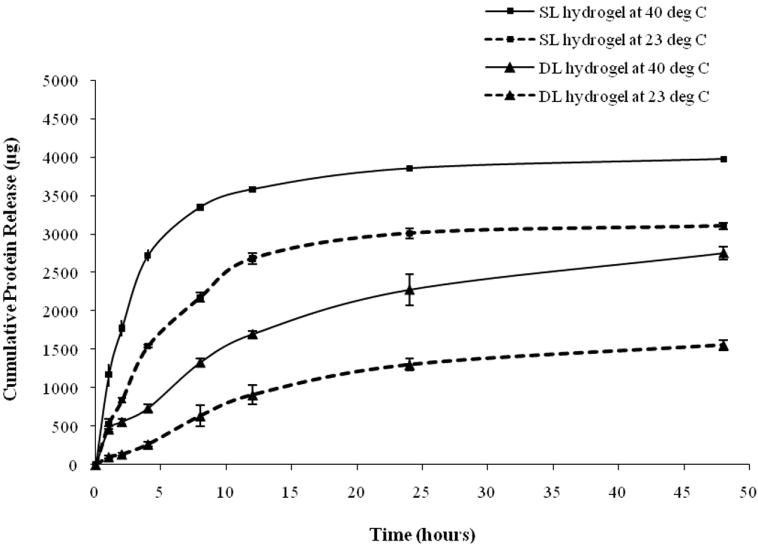
A smart protein delivery system for wound healing applications was developed using composite nanoparticle hydrogels that can release protein in a temperature-responsive manner. This system can also be formed in situ in the presence of ultraviolet light and Irgacure 2959 photoinitiator. The system consists of temperature-sensitive poly(N-isopropylacrylamide-co-acrylamide) (PNIPAM-AAm) nanoparticles embedded in a poly(ethylene glycol) diacrylate (PEGDA) matrix. A factorial analysis was performed to evaluate the effects of PEGDA concentration (10% and 15% w/v) and PEGDA molecular weight (MW; 3.4 kDa and 8 kDa), as well as PNIPAM-AAm nanoparticle concentration (2% and 4% w/v) and temperature (23 degrees C and 40 degrees C) on the protein release profiles and swelling ratios of the hydrogels. Results indicate that PNIPAM-AAm nanoparticle concentration and temperature were the most important factors affecting the protein release during the burst release phase. Additionally, PEGDA MW was the most important factor affecting the protein release in the plateau region. It was also important in controlling the hydrogel swelling ratio. A dual-layered hydrogel was further developed to produce a protein delivery system with a better sustained release. These findings have improved our understanding of the composite hydrogel systems and will help in tailoring future systems with desired release profiles.

From the clinical editor: A smart protein delivery system for wound healing applications using composite nanoparticle hydrogels that can release protein in a temperature-responsive manner is reported in this paper. Systems like this may aid in optimal would healing in the surgical and trauma-related settings.


Conflict of interest statement

There is no potential, perceived or real conflict of interest.

Figures

Figure 1
Figure 1
Principle of the photopolymerizable thermoresponsive composite nanoparticle hydrogels
Figure 2
Figure 2
(a) Comparison of identical composite nanoparticle hydrogels incubated at 23°C (left) and 40°C (right); (b) SEM image of composite nanoparticle hydrogels with 20% (w/v) PNIPAM-AAm nanoparticles
Figure 3
Figure 3
Protein release profiles for different composite hydrogels formulated as shown in Table 2. (a) Hydrogels with the molecular weight of PEGDA (M) at 3.4 kDa, the concentration of PEGDA (P) at 10% w/v, and the concentration of PNIPAM-AAm nanoparticles (N) at 2% w/v. (b) Hydrogels with M at 8 kDa, P at 10% w/v, and N at 4% w/v. (c) Hydrogels with M at 8 kDa, P at 15% w/v, and N at 2% w/v. (d) Hydrogels with M at 3.4 kDa, P at 15% w/v, and N at 4% w/v.
Figure 3
Figure 3
Protein release profiles for different composite hydrogels formulated as shown in Table 2. (a) Hydrogels with the molecular weight of PEGDA (M) at 3.4 kDa, the concentration of PEGDA (P) at 10% w/v, and the concentration of PNIPAM-AAm nanoparticles (N) at 2% w/v. (b) Hydrogels with M at 8 kDa, P at 10% w/v, and N at 4% w/v. (c) Hydrogels with M at 8 kDa, P at 15% w/v, and N at 2% w/v. (d) Hydrogels with M at 3.4 kDa, P at 15% w/v, and N at 4% w/v.
Figure 4
Figure 4
Half-normal plot showing the effect of factors on the (a) protein release rate in phase I (burst release), (c) protein release rate in phase II (sustained burst release), (e) protein release rate in phase III (plateau release), and (h) thermoresponsiveness of the hydrogels (up to 8 hrs); 3D surface plot showing the effect of factors on the (b) protein release rate in phase I (burst release), (d) protein release rate in phase II (sustained burst release), (f, g) protein release rate in phase III (plateau release), and (i) thermoresponsiveness of the hydrogels (up to 8 hrs)
Figure 4
Figure 4
Half-normal plot showing the effect of factors on the (a) protein release rate in phase I (burst release), (c) protein release rate in phase II (sustained burst release), (e) protein release rate in phase III (plateau release), and (h) thermoresponsiveness of the hydrogels (up to 8 hrs); 3D surface plot showing the effect of factors on the (b) protein release rate in phase I (burst release), (d) protein release rate in phase II (sustained burst release), (f, g) protein release rate in phase III (plateau release), and (i) thermoresponsiveness of the hydrogels (up to 8 hrs)
Figure 5
Figure 5
(a) Half-normal plot and (b) 3D surface plot showing the effect of the processing factors on the hydrogel swelling ratio
Figure 5
Figure 5
(a) Half-normal plot and (b) 3D surface plot showing the effect of the processing factors on the hydrogel swelling ratio
Figure 6
Figure 6
(a) Structure of the double layer composite nanoparticle hydrogel, (b) protein release profiles of single layer (SL) and double layer (DL) hydrogels at 23°C and 40°C
Figure 6
Figure 6
(a) Structure of the double layer composite nanoparticle hydrogel, (b) protein release profiles of single layer (SL) and double layer (DL) hydrogels at 23°C and 40°C

References

    1. Ramanan R, Chellamuthu P, Tang L, Nguyen K. Development of a temperature-sensitive composite hydrogel for drug delivery applications. Biotechnol Prog. 2006;22(1):118–125. - PubMed
    1. Quick D, Anseth K. DNA delivery from photocrosslinked PEG hydrogels: encapsulation efficiency, release profiles, and DNA quality. J Control Release. 2004;96:341–351. - PubMed
    1. Mellott MB, Searcy K, Pishko MV. Release of protein from highly cross-linked hydrogels of poly(ethylene glycol) diacrylate fabricated by UV polymerization. Biomaterials. 2001 May;22(9):929–941. - PubMed
    1. Elisseeff J, McIntosh W, Anseth K, Riley S, Ragan P, Langer R. Photoencapsulation of chondrocytes in poly (ethylene oxide)-based semi-interpenetrating networks. J Biomed Mater Res. 2000;51:164–171. - PubMed
    1. Bryant S, Anseth K. The effects of scaffold thickness on tissue engineered cartilage in photocrosslinked poly(ethylene oxide) hydrogels. Biomaterials. 2001;22:619–626. - PubMed

Publication types