. 2009 Apr 28;420(1):37-47.
doi: 10.1042/BJ20090082.

Biochemical evidence of the interactions of membrane type-1 matrix metalloproteinase (MT1-MMP) with adenine nucleotide translocator (ANT): potential implications linking proteolysis with energy metabolism in cancer cells

Affiliations

Biochemical evidence of the interactions of membrane type-1 matrix metalloproteinase (MT1-MMP) with adenine nucleotide translocator (ANT): potential implications linking proteolysis with energy metabolism in cancer cells

Ilian A Radichev et al. Biochem J. .

Abstract

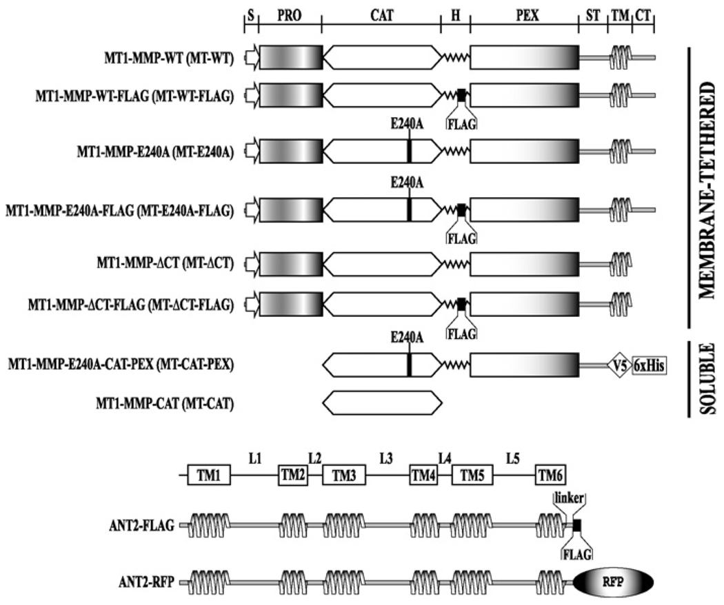
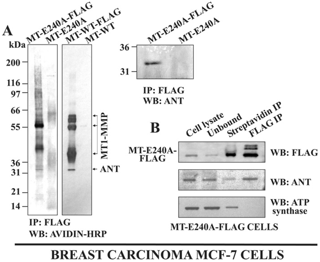
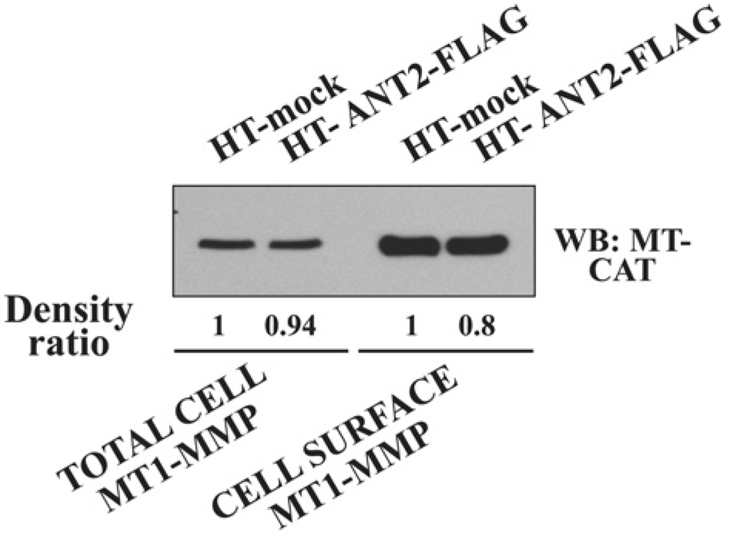
Invasion-promoting MT1-MMP (membrane type-1 matrix metalloproteinase) is a key element in cell migration processes. To identify the proteins that interact and therefore co-precipitate with this proteinase from cancer cells, we used the proteolytically active WT (wild-type), the catalytically inert E240A and the C-end truncated (tailless; DeltaCT) MT1-MMP-FLAG constructs as baits. The identity of the pulled-down proteins was determined by LC-MS/MS (liquid chromatography tandem MS) and then confirmed by Western blotting using specific antibodies. We determined that, in breast carcinoma MCF cells (MCF-7 cells), ANT (adenine nucleotide translocator) efficiently interacted with the WT, E240A and DeltaCT constructs. The WT and E240A constructs also interacted with alpha-tubulin, an essential component of clathrin-mediated endocytosis. In turn, tubulin did not co-precipitate with the DeltaCT construct because of the inefficient endocytosis of the latter, thus suggesting a high level of selectivity of our test system. To corroborate these results, we then successfully used the ANT2-FLAG construct as a bait to pull-down MT1-MMP, which was naturally produced by fibrosarcoma HT1080 cells. We determined that the presence of the functionally inert catalytic domain alone was sufficient to cause the proteinase to interact with ANT2, thus indicating that there is a non-proteolytic mode of these interactions. Overall, it is tempting to hypothesize that by interacting with pro-invasive MT1-MMP, ANT plays a yet to be identified role in a coupling mechanism between energy metabolism and pericellular proteolysis in migrating cancer cells.


Figures

Figure 1
Figure 1. Constructs of MT1-MMP and ANT2
The FLAG tag sequence was inserted in the hinge region and linked to the C-terminus of MT1-MMP and ANT2, respectively. In the ANT2-RFP chimaera, RFP was C-terminally linked to the ANT2 sequence. S, signal peptide; PRO, prodomain; CAT, catalytic domain; H, hinge region; PEX, PEX domain; ST, stalk region; TM, transmembrane domain; CT, cytoplasmic tail; L1–L5, short loop sequences that link the six transmembrane domains of ANT2.
Figure 2
Figure 2. MT1-MMP specifically interacts with ANT2 in breast carcinoma MCF cells
(A) MT1-MMP–FLAG was immunoprecipitated (IP) with anti-FLAG beads from MT-E240A–FLAG, MT-WT–FLAG, MT-ΔCT–FLAG and MT-E240A cells (control). The precipitated samples were separated by gel electrophoresis and stained with a silver stain. (B) Left panel: MT1-MMP was immunoprecipitated from MT-E240A cells using the monoclonal antibody 3G4 against the catalytic domain. The precipitated sample was separated by gel electrophoresis and stained with a silver stain. Right panel: to show the presence of ANT2, an aliquot of the precipitated sample was analysed by Western blotting (WB) with the ANT antibody. ANT2 was absent in the control sample immunoprecipitated from MT-E240A cells with anti-FLAG beads. (C) MT1-MMP-FLAG was immunoprecipitated with anti-FLAG beads from MT-E240A–FLAG, MT-WT–FLAG and MT-ΔCT–FLAG cells and also from MT-E240A, MT-WT and MT-ΔCT cells (the latter three are controls). The immunoprecipitated samples were analysed by Western blotting with the ANT and α-tubulin antibodies. The presence of the MT1-MMP–FLAG constructs in the experimental samples was confirmed using Western blotting with the FLAG antibody (upper panels).
Figure 3
Figure 3. ANT2 specifically interacts with MT1-MMP in fibrosarcoma HT cells
(A) Left panel: ANT2–FLAG was immunoprecipitated (IP) with anti-FLAG-beads from ANT2–FLAG cells and mock-transfected control cells (two left lanes). As an additional control, naturally produced MT1-MMP was immunoprecipitated from mock cells using the 3G4 antibody against MT-CAT (right lane, MT-CAT). The precipitated samples were separated by gel electrophoresis and stained with a silver stain. Right panel: the ANT2–FLAG and mock samples precipitated with anti-FLAG beads were analysed by Western blotting (WB) with the ANT and MT1-MMP 3G4 antibodies. (B) The ANT2–FLAG and mock cell lysates (cell lysates) were fractionated using anti-FLAG-beads to separate the unbound and anti-FLAG-bound (IP) protein fractions. The fractions were analysed by Western blotting with the MT1-MMP Ab815 antibody to the hinge region and also with the ATP synthase, ANT, Bcl-2 and Bax antibodies. (C) The lysate of surface-biotinylated HT-ANT2–FLAG cells (cell lysate) was fractionated using streptavidin beads to separate the unbound and streptavidin-bound (IP) protein fractions. The fractions were analysed by Western blotting with the ANT antibody.
Figure 4
Figure 4. ANT2 is expressed on the surface of breast carcinoma MCF cells
(A) Left panel: MCF-MT-E240A–FLAG, MCF-MT-WT–FLAG, MCF-MT-E240A and MCF-MT-WT cells (the latter two are controls) were surface biotinylated with membrane-impermeable biotin and then lysed. The lysates were immunoprecipitated (IP) with anti-FLAG-beads. The precipitated samples were analysed by Western blotting (WB) with Extravidin conjugated with horseradish peroxidase (avidin-HRP). Right panel: Western blotting (WB) with the ANT antibody of MCF-MT-E240A–FLAG and MCF-MT-E240A samples confirms the presence of ANT in the FLAG-immunoprecipitated sample. (B) Surface-biotinylated MCF-MT-E240A–FLAG cells were lysed (cell lysate) and the biotin-labelled proteins were precipitated using streptavidin beads. The unbound fraction was immunoprecipitated with anti-FLAG beads (FLAG IP). The cell lysate, the unbound, the steptavidin-precipitated (streptavidin IP) and the FLAG IP fractions were analysed by Western blotting with the FLAG, ANT and ATP synthase antibodies.
Figure 5
Figure 5. Co-localization of MT1-MMP and ANT2 on the surface of MCF cells
MCF-MT-WT cells were transiently transfected with the ANT2–RFP chimaera. The MCF-MT-WT/ANT2–RFP cells and MCF-MT-WT cells transiently transfected with the empty vector (control) were stained with the MT1-MMP antibody followed by an Alexa Fluor® 488-conjugated secondary antibody and the co-localization (merged) of the MT1-MMP immunoreactivity (green) with the RFP fluorescence (red) were analysed using fluorescence microscopy. Nuclei were stained with DAPI. The arrow points to the region with intense co-localization of the MT1-MMP immunoreactivity with ANT2-RFP.
Figure 6
Figure 6. The individual MT-CAT is sufficient for the ANT2 binding
Left: the MCF-MT-WT/ANT2–FLAG, MCF-ANT2–FLAG, MCF-MT-WT and MCF-mock cell lysates were immunoprecipitated (IP) with anti-FLAG beads to pull-down the cellular ANT2–FLAG–MT1-MMP complex. The presence of MT1-MMP in the precipitated samples was detected by Western blotting (WB) with the Ab815 antibody against the hinge region of MT1-MMP. Right: the soluble MT-CAT and MT-CAT-PEX constructs were each allowed to interact with ANT2–FLAG that was present in the MCF-ANT2–FLAG cell lysate. The samples were then immunoprecipitated using anti-FLAG-beads (IP). The precipitated material was analysed by Western blotting (WB) with the 3G4 antibody against MT-CAT.
Figure 7
Figure 7. ANT2 does not affect the expression of cellular MT1-MMP
The levels of total cell and cell surface-associated MT1-MMP in HT-mock and HT-ANT2–FLAG cells were determined by Western blotting (WB) with the 3G4 antibody against MT-CAT. The bands were scanned and the images were digitized. The ratio of the band density is shown at the bottom of the panel.

References

    1. Lopez-Otin C, Bond JS. Proteases: multifunctional enzymes in life and disease. J. Biol. Chem. 2008;283:30433–30437. - PMC - PubMed
    1. Seiki M, Yana I. Roles of pericellular proteolysis by membrane type-1 matrix metalloproteinase in cancer invasion and angiogenesis. Cancer Sci. 2003;94:569–574. - PMC - PubMed
    1. Wolf K, Wu YI, Liu Y, Geiger J, Tam E, Overall C, Stack MS, Friedl P. Multi-step pericellular proteolysis controls the transition from individual to collective cancer cell invasion. Nat. Cell Biol. 2007;9:893–904. - PubMed
    1. Strongin AY. Mislocalization and unconventional functions of cellular MMPs in cancer. Cancer Metastasis Rev. 2006;25:87–98. - PubMed
    1. Hotary K, Li XY, Allen E, Stevens SL, Weiss SJ. A cancer cell metalloprotease triad regulates the basement membrane transmigration program. Genes Dev. 2006;20:2673–2686. - PMC - PubMed

Publication types

Substances