

Review
doi: 10.1186/ar2528.
Epub 2009 Jan 30.
Antigen receptor signaling in the rheumatic diseases
Affiliations
- PMID: 19232064
- PMCID: PMC2688213
- DOI: 10.1186/ar2528
Item in Clipboard
Review
Antigen receptor signaling in the rheumatic diseases
Arthritis Res Ther.
2009.
Abstract
Antigen receptor signaling in lymphocytes has been clearly implicated in the pathogenesis of the rheumatic diseases. Here, we review evidence from mouse models in which B-cell and T-cell signaling machinery is perturbed as well as data from functional studies of primary human lymphocytes and recent advances in human genetics. B-cell receptor hyper-responsiveness is identified as a nearly universal characteristic of systemic lupus erythematosus in mice and humans. Impaired and enhanced T-cell receptor signaling are both associated with distinct inflammatory diseases in mice. Mechanisms by which these pathways contribute to disease in mouse models and patients are under active investigation.
Figures
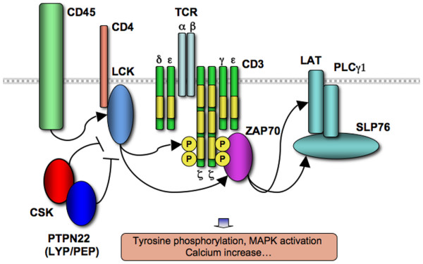
Schematic representation of T-cell receptor signal transduction. CD4-associated Lck is reciprocally regulated by CD45 and Csk/PTPN22 and in turn phosphorylates CD3 chain immunoreceptor tyrosine-based activating motifs (ITAMs) and ZAP-70. ZAP-70 phosphorylates additional downstream effectors, including the adaptors Slp-76 and Lat. Yellow bands represent CD3 chain ITAM domains. Phosphotyrosines are not depicted on all CD3 chain ITAMs. MAPK, mitogen-activated protein kinase; PLCγ1, phospholipase C γ1; TCR, T-cell antigen receptor.
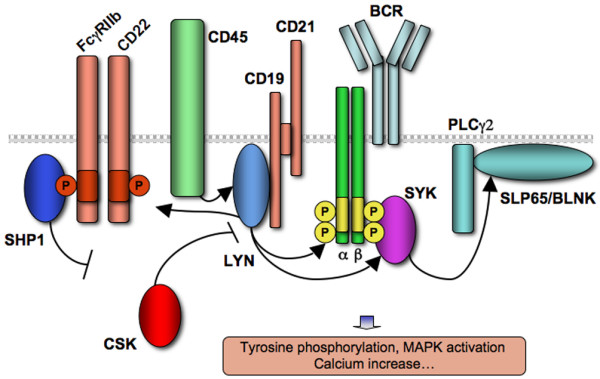
Schematic representation of B-cell receptor signal transduction. Lyn is reciprocally regulated by CD45 and Csk and in turn phosphorylates B-cell antigen receptor (BCR) immunoreceptor tyrosine-based activating motifs (ITAMs) as well as immune tyrosine inhibitory motif (ITIM)-containing immunoreceptors. Positive and negative signals are in turn transmitted by Syk and SHP-1, respectively. Yellow bands on Igα and β chains represent ITAM domains. Orange bands on CD22 and FcγRIIb represent ITIM domains. MAPK, mitogen-activated protein kinase; PLCγ2, phospholipase C γ2.
