. 2009 Feb 22:10:12.
doi: 10.1186/1471-2199-10-12.

Transcriptional inhibiton of Hoxd4 expression by miRNA-10a in human breast cancer cells

Affiliations

Transcriptional inhibiton of Hoxd4 expression by miRNA-10a in human breast cancer cells

Yuliang Tan et al. BMC Mol Biol. .

Abstract

Background: Small noncoding RNAs (ncRNAs), including short interfering RNAs (siRNAs) and microRNAs (miRNAs), can silence genes at the transcriptional, post-transcriptional or translational level 12.

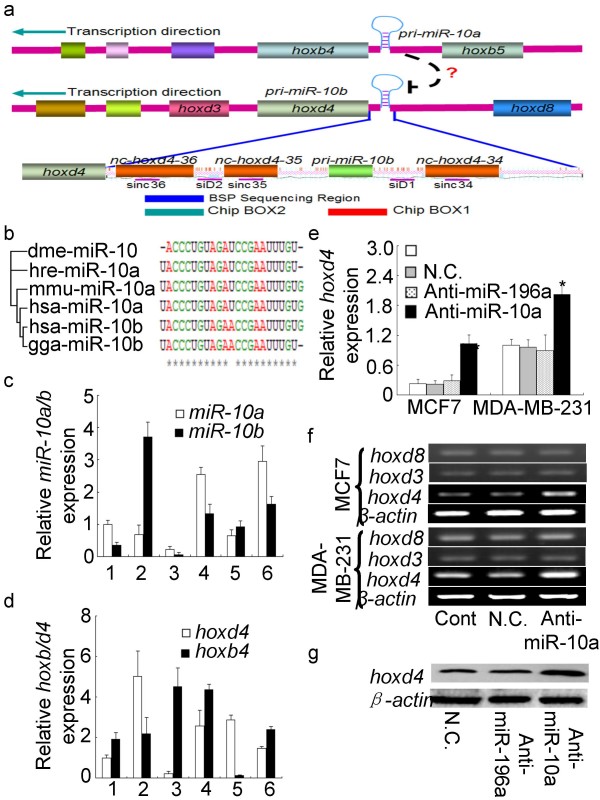
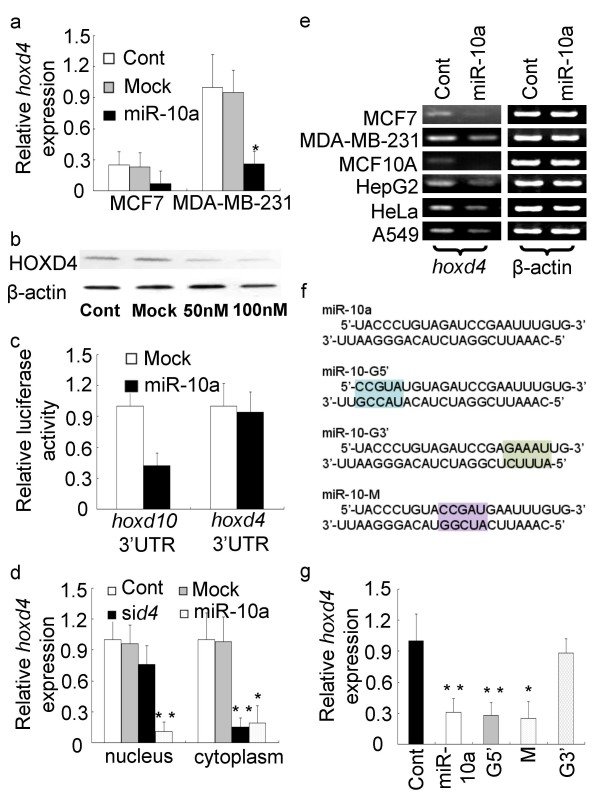
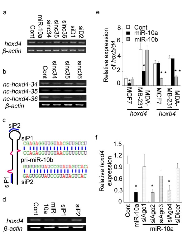
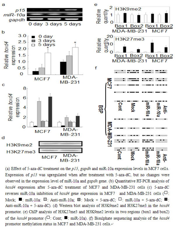
Results: Here, we show that microRNA-10a (miR-10a) targets a homologous DNA region in the promoter region of the hoxd4 gene and represses its expression at the transcriptional level. Mutational analysis of the miR-10a sequence revealed that the 3' end of the miRNA sequence is the most critical element for the silencing effect. MicroRNA-10a-induced transcriptional gene inhibition requires the presence of Dicer and Argonautes 1 and 3, and it is related to promoter associated noncoding RNAs. Bisulfite sequencing analysis showed that the reduced hoxd4 expression was accompanied by de novo DNA methylation at the hoxd4 promoter. We further demonstrated that trimethylation of histone 3 lysine 27 (H3K27me3) is involved in the miR-10a-induced hoxd4 transcriptional gene silence.

Conclusion: In conclusion, our results demonstrate that miR-10a can regulate human gene expression in a transcriptional manner, and indicate that endogenous small noncoding RNA-induced control of transcription may be a potential system for expressional regulation in human breast cancer cells.


Figures

Figure 1
Figure 1
MiR-10a-induced inhibition of hoxd4 gene expression. (a) Schematic representation of the miR-10a&b loci upstream of the hoxd4 and hoxb4 genes. The insert shows the ncRNA loci, the BSP analysed region, the siRNA target sites, and the regions analysed by ChIP in the Hoxd4 promotor. (b) MiRNA10 exhibits high evolutionary conservation in human, mouse, zebrafish, Drosophila and chicken. (c) Profile of miR-10a/b expression in cancer cells by real time PCR (1: MCF7; 2: MDA-MB-231; 3: MCF10A: 4: HepG2; 5: HeLa; 6: A549). (d) Quantitative RT-PCR analysis of hoxd4 and hoxb4 gene expression in human cancer cell lines (1: MCF7; 2: MDA-MB-231; 3: MCF10A: 4: HepG2; 5: HeLa; 6: A549). The expression miR-10a varies inversely with the expression of hoxd4 in all six cell types. (e) Quantitative PCR analysis of the hoxd4 RNA levels in MCF7 and MDA-MB-231 cells treated with antisense miR-10a 2'-O-methyl oligos, miR-196a 2'-O-methyl oligos or negative control oligos (N.C.). (f) RT-PCR analysis of the hoxd4, hoxd3 and hoxd8 mRNA levels in MCF7 and MDA-MB-231 cells treated with antisense miR-10a 2'-O-methyl oligos or negative control oligos (N.C.). (g) Protein analysis of Hoxd4 in MDA-MB-231 cells after transfection with antisense miR-10a 2'-O-methyl oligos, miR-196a 2'-O-methyl oligos or negative control oligos (N.C.).
Figure 2
Figure 2
MiR-10a modulates hoxd4 gene expression. (a) Quantitative PCR analysis of hoxd4 gene expression in MCF7 and MDA-MB-231 cells treated with miR-10a duplexes or mock oligos. (b) Protein analysis of Hoxd4 in MDA-MB-231 cells after transfection with miR-10a. When treated with 100 nM miR-10a duplexes, the expression of Hoxd4 protein was almost abolished compared to transfection with 50 nM miR-10a duplexes or control. (c) Effect of transfection of MCF7 cells with miR-10a duplexes on the relative luciferase activity of luciferase hoxd10 3'UTR and hoxd4 3'UTR reporter constructs. (d) Nuclear run-on assay of hoxd4 in the presence or absence of miR-10a and sid4. (e) RT-PCR analysis of hoxd4 expression in several human cells after transfection with miR-10a. The expression of hoxd4 mRNA was reduced after the transfection of miR-10a duplexes in all six cell types. (f) Schematic representation of the miR-10a-G5', -M and -G3' mutants. (g) Hoxd4 expression in MCF7 cells transfected with miR-10a and its mutants. The 3'most portion of miR-10a sequence is most important for miR-10a induced gene silence of hoxd4.
Figure 3
Figure 3
MiR-10a-induced transcriptional inhibition of hoxd4 is related to promoter-associated ncRNAs. (a) Effects of promoter target site on siRNA-induced inhibition of hoxd4 expression in MCF7 cells. Si-nc34 and si-nc36 inhibited hoxd4 mRNA expression in MCF7 cells, whereas siD1 and siD2 had almost no effect in hoxd4 mRNA expression. (b) RT-PCR assay of ncRNAs in the hoxd4 promotor region of MCF7 cells after transfection with si-nc34, si-nc35 and si-nc36. Si-nc35 did not affect the expression level of nc-hoxd4-35. (c) Schematic representation of the siP1 and siP2 target sites on hsa-miR-10b. SiP1 and SiP2 were designed to target the stem and lopp region, respectively, of has-miR-10b precusor. (d) Hoxd4 expression after transfection of MCF7 cells with siP1 and siP2. SiP2 couldn't affect the hoxd4 mRNA expression. (e) Expression of hoxd4 and hoxb4 (relative to β-actin) in MCF7 and MDA-MB-231 cells after transfection with miR-10a and miR-10b duplexes. MiR-10a and -10b couldn't target their own primary transcript to modulate downstream gene expression. (f) Hoxd4 expression in MCF7 cells after RNAi against Argonautes and Dicer mRNAs. The results suggested that Dicer, AGO1 and AGO3 are required for the induction of transcriptional inhibition by miR-10a duplexes.
Figure 4
Figure 4
MiR-10a-induced transcriptional gene inhibition is associated with DNA methylation of the hoxd4 promoter. (a) Effect of 5-aza-dC treatment on the p15, gapdh and miR-10a expression levels in MCF7 cells. Expression of p15 was upregulated when after treatment with 5-aza-dC, but no changes were observed in the expression level of miR-10a and gapdh gene. (b) Quantitative RT-PCR analysis of hoxd4 expression after 5-aza-dC treatment of MCF7 and MDA-MB-231 cells (c) 5-aza-dC reverses miR-10a inhibition of hoxd4 gene expression in MCF7 and MDA-MB-231 cells (□: Mock; ■: miR-10a: formula image: Anti-miR-10a; formula image: Mock + 5-aza-dC; □: miR-10a + 5-aza-dC; formula image: Anti-miR-10a + 5-aza-dC). (d) Western blot analysis of H3K9me2 and H3K27me3 in the hoxd4 promotor. (e) ChIP analysis of H3K27me3 and H3K9me2 levels in two regions (box1 and box2) of the hoxd4 promotor (□: Cont; ■: miR-10a). (f) Bisulphate sequencing analysis of the hoxd4 promotor methylation status in MCF7 and MDA-MB-231 cells.

Similar articles

Cited by

References

    1. Morris KV. RNA-mediated transcriptional gene silencing in human cells. Curr Top Microbiol Immunol. 2008;320:211–24. - PubMed
    1. Stefani G, Slack FJ. Small non-coding RNAs in animal development. Nat Rev Mol Cell Biol. 2008;9:219–30. - PubMed
    1. Bartel DP. MicroRNAs: genomics, biogenesis, mechanism, and function. Cell. 2004;116:281–97. - PubMed
    1. He L, et al. A microRNA component of the p53 tumour suppressor network. Nature. 2007;447:1130–4. - PMC - PubMed
    1. Johnson SM, et al. RAS is regulated by the let-7 microRNA family. Cell. 2005;120:635–47. - PubMed

Publication types