Three-dimensional echocardiography. New possibilities in mitral valve assessment
- PMID: 19232192
- PMCID: PMC2822664
Three-dimensional echocardiography. New possibilities in mitral valve assessment
Abstract
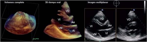
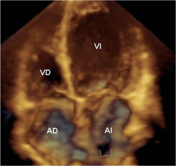
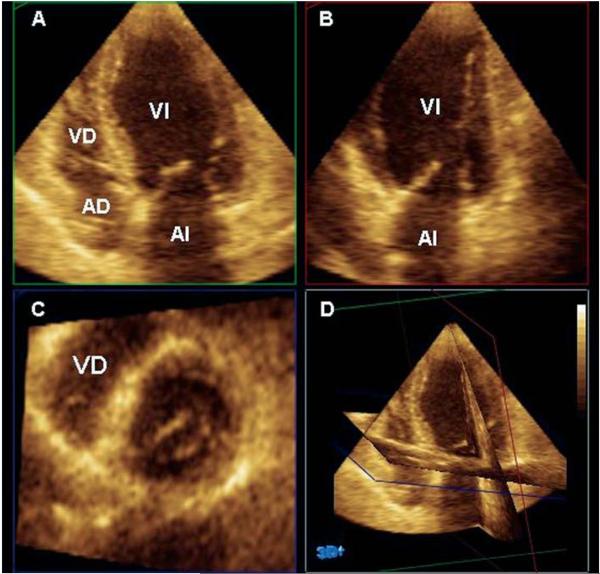
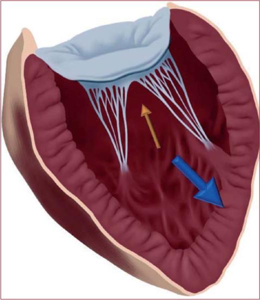
Recent developments in three-dimensional echocardiography have made it possible to obtain images in real time, without the need for off-line reconstruction. These developments have enabled the technique to become an important tool for both research and daily clinical practice. A substantial proportion of the studies carried out using three-dimensional echocardiography have focused on the mitral valve, the pathophysiology of mitral valve disease and, in particular, functional mitral regurgitation. The aims of this article were to review the contribution of three-dimensional echocardiography to understand the functional anatomy of the mitral valve and to summarize the resulting clinical applications and therapeutic implications.
Figures

References
-
- Dekker DL, Piziali RL, Dong E., Jr. A system for ultrasonically imaging the human heart in three dimensions. Comput Biomed Res. 1974;7:544–53. - PubMed
-
- Buck T, Hunold P, Wentz KU, Tkalec W, Nesser HJ, Erbel R. Tomographic three-dimensional echocardiographic determination of chamber size and systolic function in patients with left ventricular aneurysm: comparison to magnetic resonance imaging, cineventriculography, and two-dimensional echocardiography. Circulation. 1997;96:4286–97. - PubMed
-
- Jacobs LD, Salgo IS, Goonewardena S, Weinert L, Coon P, Bardo D, et al. Rapid online quantification of left ventricular volume from real-time three-dimensional echocardiographic data. Eur Heart J. 2006;27:460–8. - PubMed
-
- Ota T, Fleishman CE, Strub M, Stetten G, Ohazama CJ, Von Ramm OT, et al. Real-time, three-dimensional echocardiography: feasibility of dynamic right ventricular volume measurement with saline contrast. Am Heart J. 1999;137:958–66. - PubMed
Publication types
MeSH terms
Grants and funding
LinkOut - more resources
Full Text Sources
Medical