. 2009 Apr 3;381(2):264-70.
doi: 10.1016/j.bbrc.2009.02.035. Epub 2009 Feb 14.

Differentiation of murine embryonic stem cells to thyrocytes requires insulin and insulin-like growth factor-1

Affiliations

Differentiation of murine embryonic stem cells to thyrocytes requires insulin and insulin-like growth factor-1

Maria C Arufe et al. Biochem Biophys Res Commun. .

Abstract

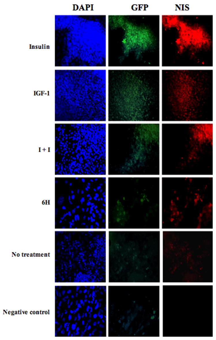
The mechanisms controlling thyrocyte development during embryonic stem (ES) cell differentiation have only been partially elucidated, although previous studies have suggested the participation of thyroid stimulating hormone (TSH) in these processes. To further define the role of TSH in this context, we have studied a murine ES cell line in which green fluorescent protein (GFP) cDNA is targeted to the TSH receptor (TSHR) gene, linking the expression of GFP to the transcription of the endogenous TSHR gene. We demonstrate that, in the initial stages of embryoid body formation, activin A and TSH induce the differentiation of definitive endoderm and thyrocyte progenitors expressing Sox17, Foxa2, and TSHR. These thyrocyte progenitors are then converted into cellular aggregates that, in the presence of insulin and IGF-1, further differentiate into mature thyroglobulin-expressing thyrocytes. Our data suggest that, despite the fact that TSH is important for the induction and specification of thyrocytes from ES cells, insulin and IGF-1 are crucial for thyrocyte maturation. Our method provides a powerful in vitro differentiation model for studying the mechanisms of early thyrocyte lineage development.


Figures

Figure 1
Figure 1. The protocol for the generation of thyrocytes from ES cells
Undifferentiated mouse ES cells were cultured on irradiated mouse embryonic fibroblasts in IMDM containing LIF. To induce EB formation, ES cells were trypsinized into a single-cell suspension and were differentiated in EBDM for 2 days to form cellular aggregates. The differentiated cells were then further induced with 10 ng/ml activin A and TSH for 4 days in KSR medium. The EB-derived cells were further cultured for 5 to 10 days in the maturation medium containing TSH, insulin and IGF-1. Abbreviation: LIF, leukemia inhibitory factor; TSH, thyroid stimulating hormone; IGF-1, insulin-growth factor-1; EBDM, embryoid body differentiation medium; KSR, knockout serum replacement medium.
Figure 2
Figure 2. Differentiation capacity of mouse TSHR+/ ES cells treated with activin A and TSH
A. RT-PCR expression analysis of various markers characteristic of endoderm and mesoderm germ layers, including Foxa2, Sox 17 and brachyury, in TSHR+/ EBs differentiated in serum-containing medium followed by serum-free medium supplemented with activin A. TSHR is a thyrocyte marker. Rex 1 is a stem cell marker. Numbers on top of the figure indicate days of differentiation. β-actin serves as an internal control. Control experiments contained no reverse transcriptase (−RT). B. Kinetics of GFP expression in early EBs. C. RT-PCR analysis of gene expression profiles in activin A-treated EBs exposed to varying concentrations of TSH. D. FACS analysis of GFP expression in EBs differentiated in serum-free medium supplemented with TSH (right panel). Control groups were not treated with TSH (left panel). E. RT-PCR analysis of gene expression profiles in day 11 and day 16 EBs treated with different conditions. Abbreviations: I+I, insulin and IGF-1; 6H, 6 hormone medium; NT, no treatment. (* indicates the band for Tg transcript).
Figure 3
Figure 3. Characterization of thyrocytes derived from mouse ES cells
Two-color immunofluorescent analysis was performed on day 11 of differentiation. Cells were stained with an antibody to NIS (red). TSHR expression is indicated by GFP (green). DAPI staining was used to identify the nucleus (blue). Note that immunofluorescence is not detected with the isotype control antibody. Abbreviations: I+I, insulin and IGF-1; 6H, 6 hormone medium.
Figure 4
Figure 4. Further characterization of thyrocytes derived from mouse ES cells
A. Microscopic analysis of cell aggregates derived from mouse TSHR+/ ES cells after 11 days of differentiation and stained with an antibody to NIS (red). TSHR expression is indicated by the GFP signal (green). An overlaid image shows the co-localization of NIS with TSHR (yellow). DAPI staining was used to identify the nucleus (blue). Note that high magnification demonstrates NIS expression in the plasma membrane. B. Immunofluorescent images of several cell clusters derived from mouse ES cells after 16 days of differentiation.

References

    1. Keller GM. In vitro differentiation of embryonic stem cells. Curr Opin Cell Biol. 1995;7:862–869. - PubMed
    1. Lin RY, Kubo A, Keller GM, Davies TF. Committing embryonic stem cells to differentiate into thyrocyte-like cells in vitro. Endocrinology. 2003;144:2644–2649. - PubMed
    1. Lin RY, Davies TF. Derivation and characterization of thyrocyte-like cells from embryonic stem cells in vitro. Methods Mol Biol. 2006;330:249–261. - PubMed
    1. Arufe MC, Lu M, Kubo A, Keller G, Davies TF, Lin RY. Directed differentiation of mouse embryonic stem cells into thyroid follicular cells. Endocrinology. 2006;147:3007–3015. - PMC - PubMed
    1. Kubo A, Shinozaki K, Shannon JM, Kouskoff V, Kennedy M, Woo S, Fehling HJ, Keller G. Development of definitive endoderm from embryonic stem cells in culture. Development. 2004;131:1651–1662. - PubMed

Publication types

MeSH terms