. 2009 Apr 10;386(2):249-56.
doi: 10.1016/j.virol.2009.01.018. Epub 2009 Feb 20.

SYNCRIP (synaptotagmin-binding, cytoplasmic RNA-interacting protein) is a host factor involved in hepatitis C virus RNA replication

Affiliations

SYNCRIP (synaptotagmin-binding, cytoplasmic RNA-interacting protein) is a host factor involved in hepatitis C virus RNA replication

Helene Minyi Liu et al. Virology. .

Abstract

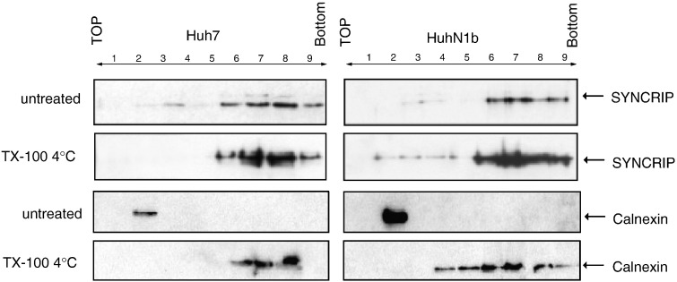
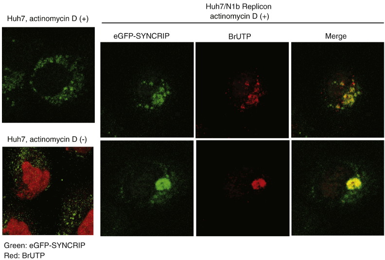
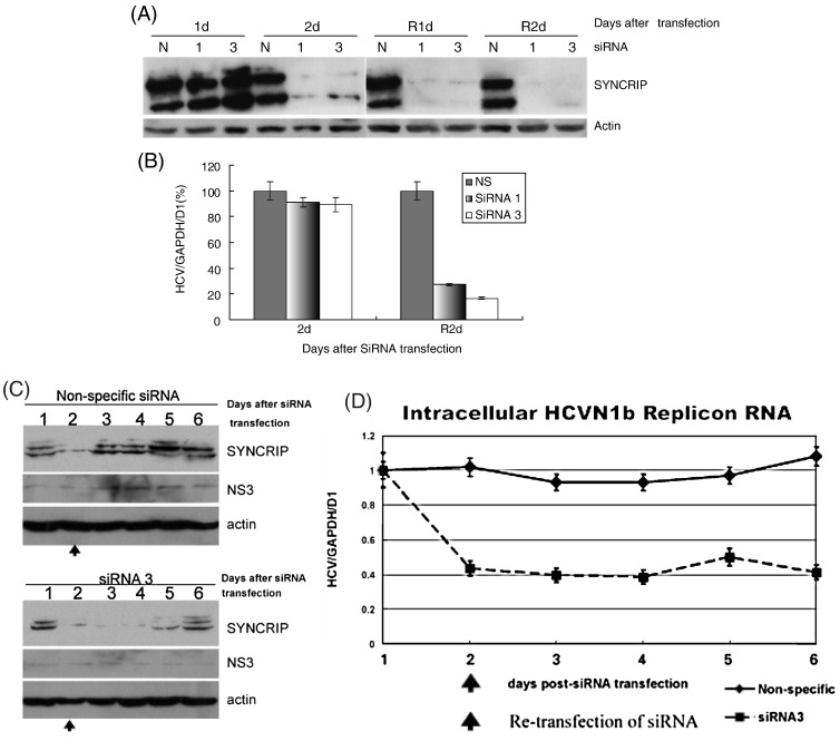
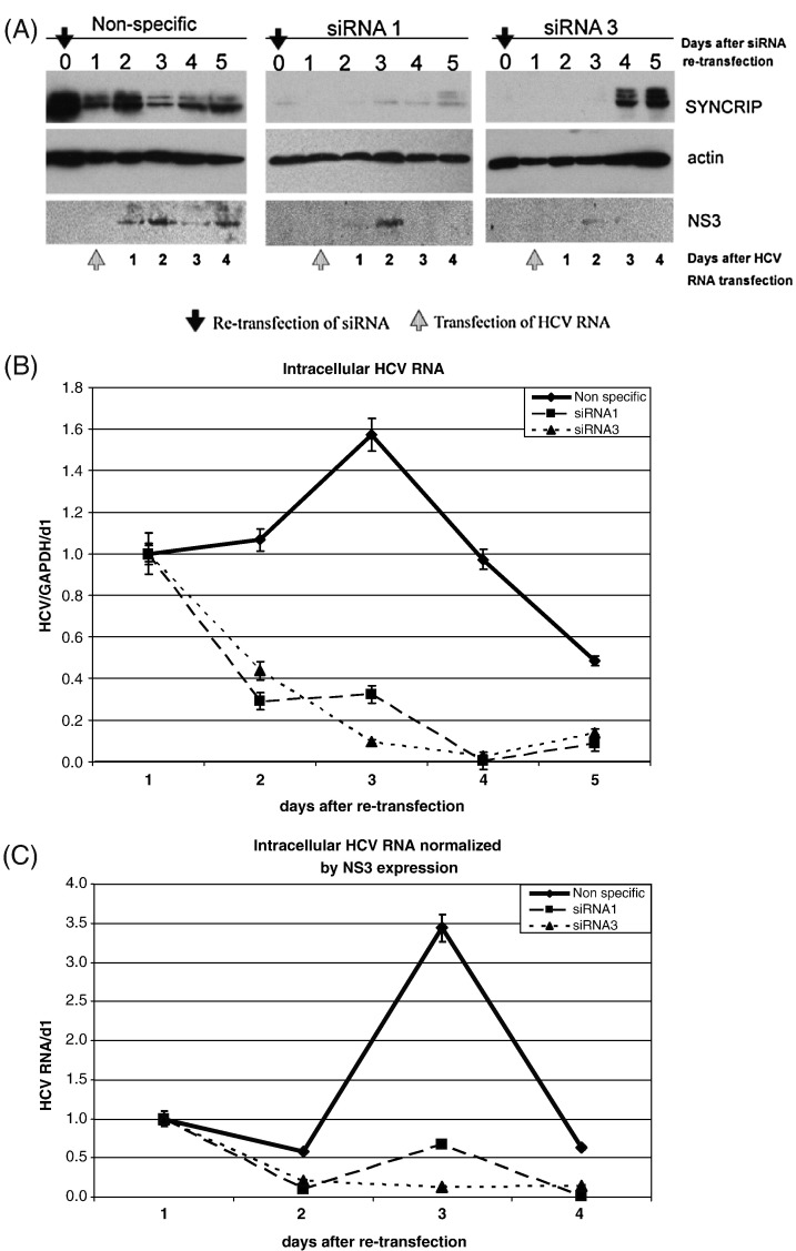
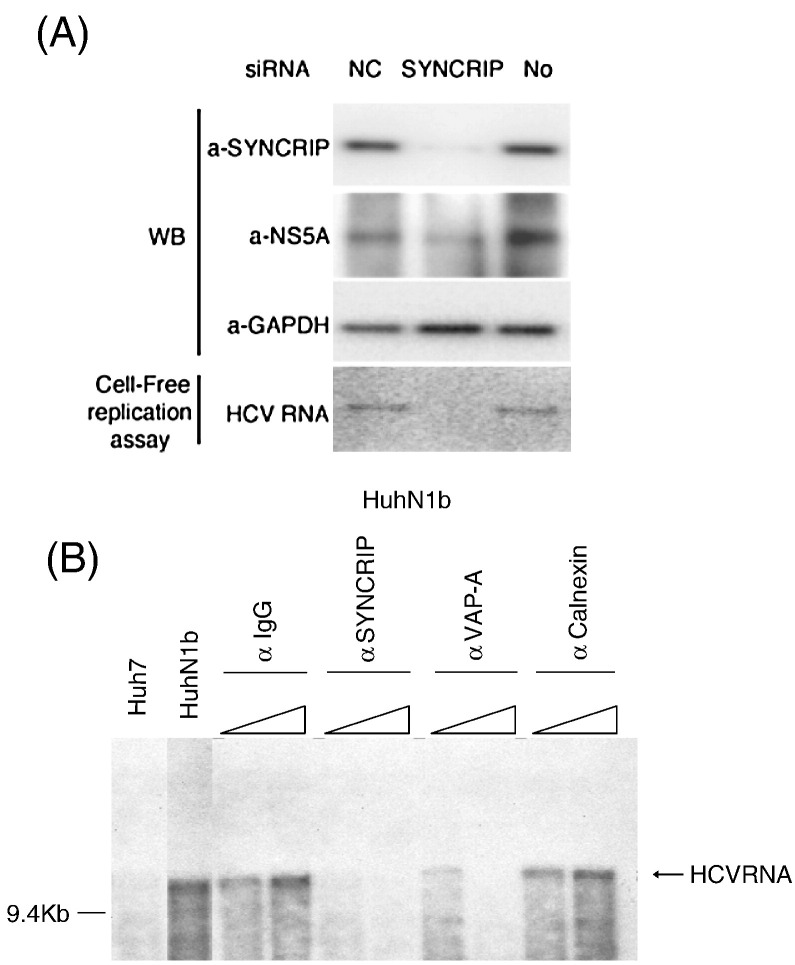
Hepatitis C virus (HCV) RNA replication requires viral nonstructural proteins as well as cellular factors. Recently, a cellular protein, synaptotagmin-binding, cytoplasmic RNA-interacting protein (SYNCRIP), also known as NSAP1, was found to bind HCV RNA and enhance HCV IRES-dependent translation. We investigate whether this protein is also involved in the HCV RNA replication. We found that SYNCRIP was associated with detergent-resistant membrane fractions and colocalized with newly-synthesized HCV RNA. Knock-down of SYNCRIP by siRNA significantly decreased the amount of HCV RNA in the cells containing a subgenomic replicon or a full-length viral RNA. Lastly, an in vitro replication assay after immunodepletion of SYNCRIP showed that SYNCRIP was directly involved in HCV RNA replication. These findings indicate that SYNCRIP has dual functions, participating in both RNA replication and translation in HCV life cycle.


Figures

Fig. 1
Fig. 1
Membrane flotation assay showed relocalization of SYNCRIP to DRM in HCV replicon cells. Cell lysates were prepared from HCV replicon (HuhN1b) or Huh7 cells by passing through a 25-gauge needle 20 times. Nuclei and unbroken cells were removed by centrifugation at 1000 ×g for 5 min in microcentrifuge at 4 °C. The supernatants treated with or without 1% TX-100 at 4 °C for 30 min were fractionated by discontinuous sucrose gradient centrifugation. Fractions were collected from the top, numbered from 1 to 9. Each fraction was concentrated by Centricon YM-30 (Millipore, MA) and immunoblotted by rabbit anti-SYNCRIP antibody or mouse anti-Calnexin antibody, respectively. SYNCRIP was found in both membrane and soluble fractions in the untreated Huh7 and HuhN1b cells, whereas in the HuhN1b cells, some SYNCRIP was localized to the DRM fractions. This phenomenon is not seen in the Calnexin profile.
Fig. 2
Fig. 2
SYNCRIP colocalization with de novo-synthesized HCV RNA in a HCV replicon cell. peGFP-SYNCRIP was transfected into Huh7 or HCV replicon (HuhN1b) cells by Fugene 6. Two days after transfection, Huh7 or HCV replicon cells were labeled with BrUTP for 15 min after one-hour treatment with Actinomycin D. Actinomycin D treatment inhibited BrUTP incorporation in Huh7 cells (left two panels), but not in HuhN1b replicon cells, where BrU label was detected in the cytoplasm (right 6 panels). Immunofluorescence staining was performed with sheep polyclonal antibody against BrdU (anti-BrdU) followed by Rhodamine-conjugated anti-sheep antibody (Jackson ImmunoResearch). Two different HuhN1b cells are shown, representing two different distribution patterns of BrUTP, as shown previously (Mizutani et al., 2000).
Fig. 3
Fig. 3
SYNCRIP knock-down by siRNA in HCV replicon cells. The siRNAs against SYNCRIP (siRNA 1 and 3) (Choi et al., 2004b) or nonspecific siRNA were transfected into HCV replicon cells. Two days after the first transfection, each siRNA was re-transfected into the same cells to ensure complete knock-down of SYNCRIP. Endogenous SYNCRIP protein levels were monitored by immunoblotting (A), and HCV RNA levels were detected by realtime RT-PCR (B). (C), immunoblotting of SYNCRIP and NS3 expression in SYNCRIP siRNA knock-down replicon cells. (D), intracellular replicon RNA level was examined by realtime RT-PCR. R1d and R2d, one or 2 days after re-transfection of siRNA. siRNA 1 and 3, two different clones of siRNA; N, nonspecific siRNA.
Fig. 4
Fig. 4
Deficiency of full-length HCV replication in SYNCRIP knock-down Huh7 cells. The siRNAs against SYNCRIP (siRNA 1 and 3) or nonspecific siRNA were transfected into Huh7 cells twice to knock-down endogenous SYNCRIP level as described in Fig. 3. One day after re-transfection of siRNA, in vitro synthesized full-length HCV-1b-hyb RNA was transfected to siRNA-transfected Huh7 cells respectively. (A), immunoblotting of endogenous SYNCRIP after siRNA re-transfection, and NS3 expression levels at various days after HCV RNA transfection. (B), intracellular HCV RNA levels determined by quantitative RT-PCR, and (C), intracellular HCV RNA levels normalized by NS3 expression levels. Huh7 cells were transfected with SYNCRIP-specific or nonspecific siRNA as in (A). One day after siRNA transfection, the replication-competent full-length HCV RNA was transfected into the cells, and HCV RNA levels were detected by realtime RT-PCR on different days after the HCV RNA transfection. The relative amounts of HCV RNA are expressed as in Fig. 3B.
Fig. 5
Fig. 5
siRNA-knock-down or immunodepletion of SYNCRIP inhibited HCV replication activity in in vitro replication assay. (A) SYNCRIP was knocked down by transfection two times with specific siRNA in HCV replicon cells as in Fig. 3. SYNCRIP, NS5A and GAPDH protein expression were determined by immunoblotting. Cell-free RNA replication assays using the DRM fraction isolated from the cell lysates, as described in Material and methods, were performed. 32P-CTP-labeled HCV RNA product was detected by autoradiography after separation by agarose gel electrophoresis. NC, nonspecific siRNA control; SYNCRIP, SYNCRIP-specific siRNA; No, no siRNA transfection. (B) Partially purified lysates of HCV replicon cells were incubated with anti-IgG, anti-SYNCRIP, anti-VAP-A (VAP-33), or anti-Calnexin antibodies. Then samples were incubated with α-32P-CTP in a cell-free RNA-dependent RNA polymerase assay. The RNA product was separated by formaldehyde agarose gels and identified by autoradiography.

References

    1. Aizaki H., Lee K.J., Sung V.M., Ishiko H., Lai M.M. Characterization of the hepatitis C virus RNA replication complex associated with lipid rafts. Virology. 2004;324(2):450–461. - PubMed
    1. Aizaki H., Choi K.S., Liu M., Li Y.J., Lai M.M. Polypyrimidine-tract-binding protein is a component of the HCV RNA replication complex and necessary for RNA synthesis. J. Biomed. Sci. 2006;13(4):469–480. - PubMed
    1. Ali N., Siddiqui A. Interaction of polypyrimidine tract-binding protein with the 5′ noncoding region of the hepatitis C virus RNA genome and its functional requirement in internal initiation of translation. J. Virol. 1995;69(10):6367–6375. - PMC - PubMed
    1. Ali N., Siddiqui A. The La antigen binds 5′ noncoding region of the hepatitis C virus RNA in the context of the initiator AUG codon and stimulates internal ribosome entry site-mediated translation. Proc. Natl. Acad. Sci. U. S. A. 1997;94(6):2249–2254. - PMC - PubMed
    1. Ali N., Tardif K.D., Siddiqui A. Cell-free replication of the hepatitis C virus subgenomic replicon. J. Virol. 2002;76(23):12001–12007. - PMC - PubMed

MeSH terms

Substances