. 2009 Apr;37(7):2204-10.
doi: 10.1093/nar/gkp086. Epub 2009 Feb 20.

Methylation-state-specific recognition of histones by the MBT repeat protein L3MBTL2

Affiliations

Methylation-state-specific recognition of histones by the MBT repeat protein L3MBTL2

Yahong Guo et al. Nucleic Acids Res. 2009 Apr.

Abstract

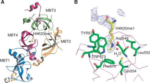
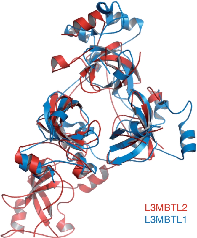
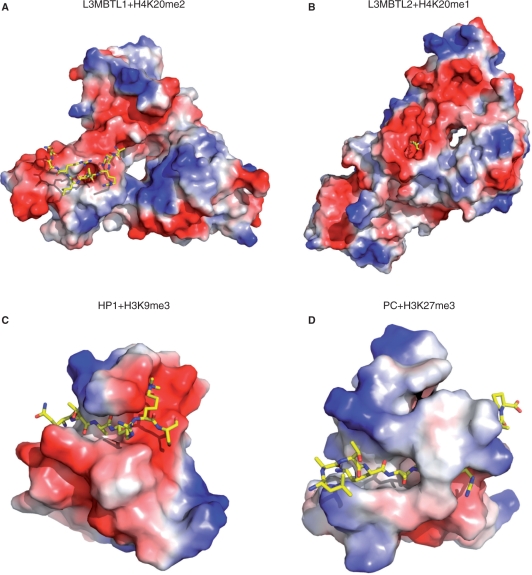
The MBT repeat has been recently identified as a key domain capable of methyl-lysine histone recognition. Functional work has pointed to a role for MBT domain-containing proteins in transcriptional repression of developmental control genes such as Hox genes. In this study, L3MBTL2, a human homolog of Drosophila Sfmbt critical for Hox gene silencing, is demonstrated to preferentially recognize lower methylation states of several histone-derived peptides through its fourth MBT repeat. High-resolution crystallographic analysis of the four MBT repeats of this protein reveals its unique asymmetric rhomboid architecture, as well as binding mechanism, which preclude the interaction of the first three MBT repeats with methylated peptides. Structural elucidation of an L3MBTL2-H4K20me1 complex and comparison with other MBT-histone peptide complexes also suggests that an absence of distinct surface contours surrounding the methyl-lysine-binding pocket may underlie the lack of sequence specificity observed for members of this protein family.


Figures

Figure 1.
Figure 1.
Isothermal titration calorimetry data for binding of unmodified and different methylated H4K20 peptides (AKRHRK20VLRDN in sequence) to the 4-MBT-repeat domain (4MBT) of L3MBTL2. ND: No detectable binding.
Figure 2.
Figure 2.
Only the fourth MBT repeat of L3MBTL2 binds H4K20me1. (A) Overall structure of 4MBT bound to an H4K20me1 peptide (AKRHRK20VLRDN). MBT1, MBT2, MBT3 and MBT4 are shown in blue, yellow, green and purple, respectively. The K20me1 lysine is shown in a stick model. (B) K20me1 recognition by the fourth repeat MBT4. The lysine-binding pocket residues and K20me1 are shown in a stick model. The electron density map 2Fo–Fc is contoured at 1 σ. The electron density map is calculated with the peptide omitted.
Figure 3.
Figure 3.
Superposition of the crystal structures of L3MBTL1 and L3MBTL2 MBT domains. L3MBTL1 is colored in blue and L3MBTL2 is colored in red.
Figure 4.
Figure 4.
L3MBTL2 uses a cavity insertion recognition mode to recognize methyl–lysine histones. (A) Surface representation of the L3MBTL1 and H4K20me2 complex structure (PDB 2PQW). (B) Surface representation of the L3MBTL2 and H4K20me1 complex structure (PDB 3F70). (C) Surface representation of the HP1 and H3K9me3 complex structure (PDB 1KNE). (D) Surface representation of the Pc and H3K27me3 complex structure (PDB 1PFB). Peptides are shown in a stick model.
Figure 5.
Figure 5.
Superposition of the structures of all solved peptide-bound MBT repeats viewed from two different directions (5A and 5B). L3MBTL1–H4K20me2 complex is colored in red (PDB 2PQW); L3MBTL1–H1.5 complex is colored in yellow (PDB 2RHI); Scm and RKmeS peptide complex is colored in blue (PDB 2R5M); and L3MBTL2–H4K20me1 complex is colored in green (this study). Lysine residues are shown in a stick model in (A).

References

    1. Grewal SI, Jia S. Heterochromatin revisited. Nat. Rev. 2007;8:35–46. - PubMed
    1. Cao R, Zhang Y. The functions of E(Z)/EZH2-mediated methylation of lysine 27 in histone H3. Curr. Opin. Gen. Dev. 2004;14:155–164. - PubMed
    1. Fischle W, Wang Y, Jacobs SA, Kim Y, Allis CD, Khorasanizadeh S. Molecular basis for the discrimination of repressive methyl-lysine marks in histone H3 by Polycomb and HP1 chromodomains. Genes Dev. 2003;17:1870–1881. - PMC - PubMed
    1. Jacobs SA, Khorasanizadeh S. Structure of HP1 chromodomain bound to a lysine 9-methylated histone H3 tail. Science. 2002;295:2080–2083. - PubMed
    1. Min J, Zhang Y, Xu RM. Structural basis for specific binding of Polycomb chromodomain to histone H3 methylated at Lys 27. Genes Dev. 2003;17:1823–1828. - PMC - PubMed

Publication types