. 2009 Apr;191(8):2493-500.
doi: 10.1128/JB.00027-09. Epub 2009 Feb 20.

The tubulin-like RepX protein encoded by the pXO1 plasmid forms polymers in vivo in Bacillus anthracis

Affiliations

The tubulin-like RepX protein encoded by the pXO1 plasmid forms polymers in vivo in Bacillus anthracis

Parvez Akhtar et al. J Bacteriol. 2009 Apr.

Abstract

Bacillus anthracis contains two megaplasmids, pXO1 and pXO2, that are critical for its pathogenesis. Stable inheritance of pXO1 in B. anthracis is dependent upon the tubulin/FtsZ-like RepX protein encoded by this plasmid. Previously, we have shown that RepX undergoes GTP-dependent polymerization in vitro. However, the polymerization properties and localization pattern of RepX in vivo are not known. Here, we utilize a RepX-green fluorescent protein (GFP) fusion to show that RepX forms foci and three distinct forms of polymeric structures in B. anthracis in vivo, namely straight, curved, and helical filaments. Polymerization of RepX-GFP as well as the nature of polymers formed were dependent upon concentration of the protein inside the B. anthracis cells. RepX predominantly localized as polymers that were parallel to the length of the cell. RepX also formed polymers in Escherichia coli in the absence of other pXO1-encoded products, showing that in vivo polymerization is an inherent property of the protein and does not require either the pXO1 plasmid or proteins unique to B. anthracis. Overexpression of RepX did not affect the cell morphology of B. anthracis cells, whereas it drastically distorted the cell morphology of E. coli host cells. We discuss the significance of our observations in view of the plasmid-specific functions that have been proposed for RepX and related proteins encoded by several megaplasmids found in members of the Bacillus cereus group of bacteria.


Figures

FIG. 1.
FIG. 1.
Expression of native RepX and RepX-GFP in B. anthracis. (A) Western blot analysis of RepX and RepX-GFP in cell lysates from B. anthracis (Ba) containing either the wt pXO1 plasmid encoding RepX or the multicopy pKL2509 plasmid constitutively expressing RepX-GFP. (B) Xylose (Xyl) concentration-dependent expression of RepX-GFP from pKL2468 in B. anthracis cells. Total protein from plasmid-free (Ba) and pXO1-containing B. anthracis cells was used as the control.
FIG. 2.
FIG. 2.
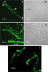
RepX-GFP assembles into polymers in B. anthracis. Fluorescence microscopic (A) and corresponding phase contrast images (B) of B. anthracis expressing RepX-GFP from pKL2509 showing straight (arrowheads) and curved (arrows) long polymers. Fluorescence microscopic (C) and corresponding phase contrast images (D) of B. anthracis harboring pKL2509 showing foci and long polymers. RepX-GFP foci at a higher magnification are shown in the inset of panel C. Fluorescence microscopic image of B. anthracis harboring pKL2509 (E), showing helical filaments assembled from RepX-GFP.
FIG. 3.
FIG. 3.
RepX-GFP requires a critical concentration of the protein for assembly of polymers in B. anthracis. (A) Western blot analysis of cell lysates derived from a B. anthracis (Ba) strain containing both the wt pXO1 plasmid expressing native RepX and the pKL2468 plasmid expressing the RepX-GFP protein. Cells were treated with various concentrations of xylose (Xyl), and affinity-purified polyclonal anti-RepX antibodies were used for Western blot analysis. Shown also is fluorescence microscopy of B. anthracis harboring only the pKL2468 plasmid treated with 0.01% (B), 0.02% (C), 0.03% (D), and 0.10% (E) concentrations of xylose as well as fluorescence microscopic images of B. anthracis cells harboring both pKL2468 and wt pXO1 after growth in 0.01% (F), 0.02% (G), and 0.03% (H) xylose. RepX-GFP foci at a higher magnification are shown in the inset of panel C.
FIG. 4.
FIG. 4.
RepX-GFP assembles into long polymers in E. coli and affects host cell phenotype. (A) Western blot analysis of RepX-GFP in B. anthracis (Ba) or E. coli (Ec) cell lysates containing either the pKL2509 plasmid expressing RepX-GFP from the native repX promoter or from the leaky Pspac promoter in the pKL2427 plasmid. Cell lysates from B. anthracis strains either lacking any plasmid (Ba) or containing the pXO1 plasmid expressing the native RepX protein were used as controls. Shown also are fluorescence microscopic (B) and corresponding phase contrast (C) images of E. coli cells harboring pKL2509 that expresses RepX-GFP as well as fluorescence microscopic (D) and corresponding phase contrast (E) images of E. coli cells harboring the pKL2427 plasmid that overexpresses RepX-GFP.

References

    1. Anand, S. P., P. Akhtar, E. Tinsley, S. C. Watkins, and S. A. Khan. 2008. GTP-dependent polymerization of the tubulin-like RepX replication protein encoded by the pXO1 plasmid of Bacillus anthracis. Mol. Microbiol. 67881-890. - PubMed
    1. Barillà, D., E. Carmelo, and F. Hayes. 2007. The tail of the ParG DNA segregation protein remodels ParF polymers and enhances ATP hydrolysis via an arginine finger-like motif. Proc. Natl. Acad. Sci. USA 1041811-1816. - PMC - PubMed
    1. Barillà, D., M. F. Rosenberg, U. Nobbmann, and F. Hayes. 2005. Bacterial DNA segregation dynamics mediated by the polymerizing protein ParF. EMBO J. 241453-1464. - PMC - PubMed
    1. Becker, E., N. C. Herrera, F. Q. Gunderson, A. I. Derman, A. L. Dance, J. Sims, R. A. Larsen, and J. Pogliano. 2006. DNA segregation by the bacterial actin AlfA during Bacillus subtilis growth and development. EMBO J. 255919-5931. - PMC - PubMed
    1. Biteen, J. S., M. A. Thompson, N. K. Tselentis, G. R. Bowman, L. Shapiro, and W. E. Moerner. 2008. Super-resolution imaging in live Caulobacter crescentus cells using photoswitchable EYFP. Nat. Methods 5947-949. - PMC - PubMed

MeSH terms

LinkOut - more resources