. 2009 Mar;136(6):923-32.
doi: 10.1242/dev.031815.

An essential role for the RNA-binding protein Smaug during the Drosophila maternal-to-zygotic transition

Affiliations

An essential role for the RNA-binding protein Smaug during the Drosophila maternal-to-zygotic transition

Beatrice Benoit et al. Development. 2009 Mar.

Abstract

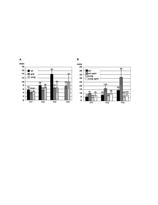
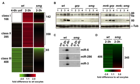
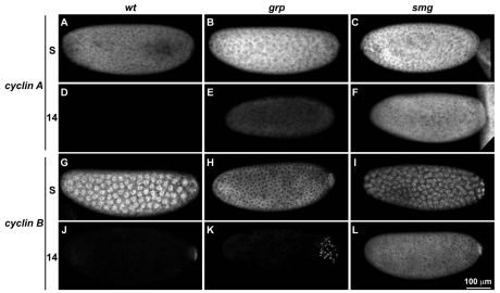
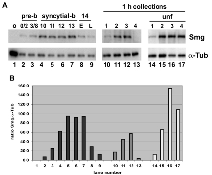
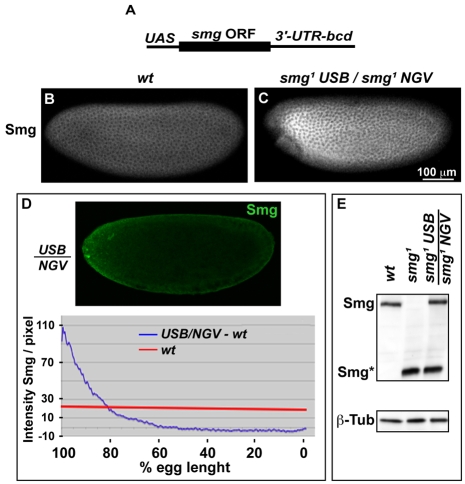
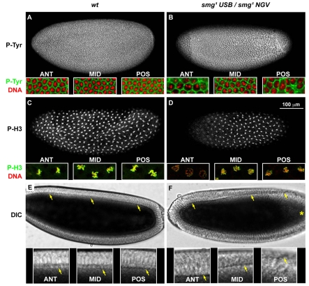
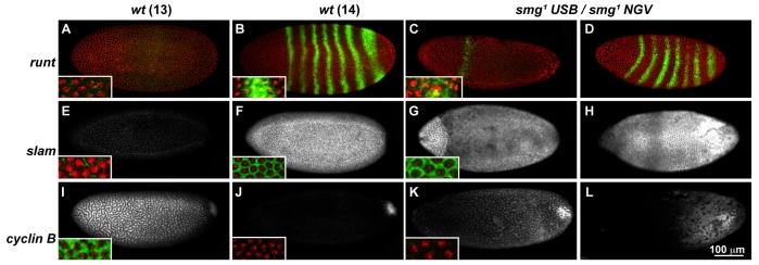
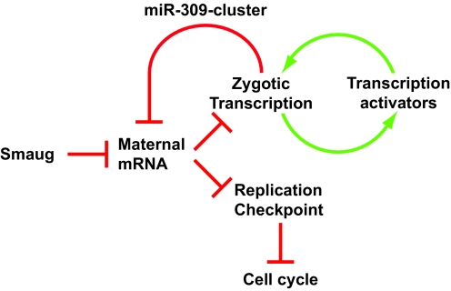
Genetic control of embryogenesis switches from the maternal to the zygotic genome during the maternal-to-zygotic transition (MZT), when maternal mRNAs are destroyed, high-level zygotic transcription is initiated, the replication checkpoint is activated and the cell cycle slows. The midblastula transition (MBT) is the first morphological event that requires zygotic gene expression. The Drosophila MBT is marked by blastoderm cellularization and follows 13 cleavage-stage divisions. The RNA-binding protein Smaug is required for cleavage-independent maternal transcript destruction during the Drosophila MZT. Here, we show that smaug mutants also disrupt syncytial blastoderm stage cell-cycle delays, DNA replication checkpoint activation, cellularization, and high-level zygotic expression of protein coding and micro RNA genes. We also show that Smaug protein levels increase through the cleavage divisions and peak when the checkpoint is activated and zygotic transcription initiates, and that transgenic expression of Smaug in an anterior-to-posterior gradient produces a concomitant gradient in the timing of maternal transcript destruction, cleavage cell cycle delays, zygotic gene transcription, cellularization and gastrulation. Smaug accumulation thus coordinates progression through the MZT.


Figures

Fig. 1.
Fig. 1.
Replication checkpoint activation during the MZT. The DNA replication checkpoint is required for increases in interphase length during syncytial blastoderm divisions 11-13. (A) Interphase length was assayed by injection of rhodamine-conjugated Tubulin and time-lapse confocal imaging. Embryos mutant for smg, like grp replication checkpoint mutants, do not show increases in interphase length during the syncytial blastoderm divisions. (B) Replication checkpoint function was assayed by co-injection of rhodamine-Tubulin and aphidicolin or carrier control. In wild-type embryos, aphidicolin induced progressively longer interphase delays during cycles 11-13. By contrast, smg mutants showed only minimal interphase delays in response to aphidicolin, and the delays did not increase with division cycle number. Each bar represents the mean interphase length (in minutes) with standard deviations. The number of individual embryos scored is given in brackets.
Fig. 2.
Fig. 2.
Smaug is required for zygotic transcription during the MZT. (A) Microarray analysis of zygotic gene expression in 2- to 3-hour-old wild-type (wt) and smg mutant embryos. Expression is relative to mature, stage 14 oocytes, which contain the full maternal pool of mRNA. Maternal genes that are not transcribed at the MZT represent ∼80% of transcripts in early embryos, and are not shown. Class I zygotic genes are not present in oocytes (not colored) and show high levels of expression in 2- to 3-hour-old embryos. One-hundred and forty-two of the 166 Class I genes required Smaug for zygotic expression. Class II genes produce maternal transcripts that are stable in unfertilized eggs and show significantly increased expression in 2- to 3-hour-old post-fertilization embryos. Three-hundred and fifty-eight of 395 Class-II genes require Smaug for zygotic expression. Class III genes produce maternal transcripts that are degraded and then re-expressed in 2-3 hour post-fertilization embryos. Sixty-five of 408 Class III genes require Smaug for expression. (B) Serine 2 phosphorylation of the RNA polymerase II CTD is linked to active transcription. Western blots reveal a dramatic increase in ser 2 phosphorylation between 2 and 4 hours of embryogenesis in wild type (wt) and mnk grp, but not in grp, smg or mnk; smg mutants. For each genotype, lane 1 shows 0- to 1-hour-old embryos, lane 2 shows 1- to 2-hour-old embryos, lane 3 shows 2- to 3-hour-old embryos and lane 4 shows 3- to 4-hour-old embryos. For each genotype, the top panel shows the hyperphosphorylated (II0, Ser2-P) and the hypophosphorylated (IIa) forms, detected with an anti-RNA polII (ARNA3). The middle panel shows phosphorylation on Ser2 (II0) detected using the phospho-epitope specific H5 antibody. The bottom panel shows α-Tubulin, which was used as a loading control. (C) Northern blots for miR-6, miR-286 and miR-3 revealed a large increase in wild-type 2- to 4-hour-old embryos, whereas expression was not detected smg mutants. (D) Heat map showing the behavior of 406 of the 410 miR-309-dependent maternal mRNAs (from Bushati et al., 2008) in embryos from wild-type and smg-mutant females relative to wild-type stage 14 oocyte reference RNA. These transcripts are unstable (green) in wild type, whereas almost 85% are stabilized in smg mutants.
Fig. 3.
Fig. 3.
Maternal mRNA degradation is independent of the replication checkpoint. Embryos mutant for smg are defective in replication checkpoint activation and maternal transcript destruction. (A-L)To determine whether checkpoint defects lead to a block in maternal transcript destruction, grp checkpoint mutant embryos were assayed for maternal cyclin A (A-F) and cyclin B (G-L) mRNA expression by whole-mount in situ hybridization. In wild-type controls, both transcripts are expressed in syncytial blastoderm embryos (S; A,G) and are degraded during interphase of cycle 14 (14 D,J). The smg mutation blocks destruction of these transcripts (C,F,I,L), but the grp mutations does not (B,E,H,K). Embryos are orientated with their anterior pole facing leftwards. S, syncytial blastoderm; 14, cycle 14. Scale bar: 100 μm.
Fig. 4.
Fig. 4.
Smaug protein expression during early embryogenesis. (A) Smaug protein expression in wild-type ovaries and embryos. Lane 1, ovary (o); lanes 2-9, embryos were fluorescently labeled, hand selected for specific division cycles (cycles indicated above lane) and subjected to western blotting (E and L indicate early and late interphase 1); lanes 10-17, western blots of pooled embryos aged 0 to 1 hour (1), 1 to 2 hours (2), 2 to 3 hours (3) and 3 to 4 hours (4); lanes 10-13 are fertilized embryos; lanes 14-17 are activated unfertilized eggs (unf). α-Tubulin is used as a loading control. (B) Quantification of Smaug expression relative to α-Tubulin for each of the lanes shown in A. In fertilized embryos, Smaug protein levels increase progressively through the early cleavage divisions, peak during cycles 11-13, and decline rapidly during interphase 14. In unfertilized eggs, the protein accumulates rapidly through the first 3 hours post egg deposition, and persists for 4 hours (lanes 14-17).
Fig. 5.
Fig. 5.
Generating a Smaug protein gradient. (A) Schematic representation of the UAS-smg-bcd transgene (USB). The yeast upstream activator sequence (UAS), Smaug coding sequence (Smaug ORF, bold line) and bcd 3′ UTR (3-UTR-bcd) are indicated. (B,C) Immunolocalization of Smaug protein in embryos derived from wild-type (wt) and smg females expressing the USB transgene. Some of the immunolabeling in the latter is due to antibody recognition of a truncated from of Smaug expressed in the smg1 mutants (see E). Embryos are oriented with their anterior pole facing leftwards. (D) Single confocal mid-section showing Smaug immunolabeling in an embryo from a wild-type female expressing the USB transgene. Average cortical pixel intensity in wild-type embryos (red line, n=4) was subtracted from average cortical pixel intensity in embryos from wild-type females expressing USB (blue line, n=4). 100% designates the anterior pole and 0% represents the posterior pole. (E) Western-blot showing Smaug protein expression in embryos from wild type (wt), smg-mutant females and smg-mutant females expressing the USB transgene. Embryos were 0-3 hours old. Full-length Smaug (Smg) and a truncated form of the protein (Smg*) expressed in smg1 mutants are indicated. β-Tubulin (β-Tub) is a loading control.
Fig. 6.
Fig. 6.
Smaug protein gradient triggers graded cell cycle delays and cellularization. (A,B) Embryos co-stained with a phospho-Tyrosine antibody (grayscale and green) and TOTO3 (red), to visualize membranes and nuclei at cellularization. (A) Nuclear density is uniform along the anterior-posterior axis of wild-type interphase 14 embryos undergoing cellularization. (B) An embryo expressing a Smaug gradient. The anterior pole cellularizes at cell cycle 13 nuclear density (ANT inset), the middle cellularizes at cycle-14 density (MID inset) and the posterior pole is disorganized (POS inset). (C,D) Embryos labeled for mitotic nuclei with anti-phospho-Histone-3 antibody (grayscale and green) and for DNA with TOTO3 (red). Wild-type cycle 11 embryos divide synchronously along the anterior-posterior axis (C). Embryos expressing Smaug in a gradient show cell cycle delays at the anterior pole (D). In this example, the posterior is in mitosis while the anterior is in late interphase or prophase (D). (E,F). Time-lapse DIC microscopy of cellularization in wild-type and USB embryos. (E) Wild-type embryos consistently cellularize synchronously along the anterior-posterior axis (see Movie 1 in the supplementary material). (F) USB embryos, by contrast, consistently initiate cellularization at the anterior pole, and membrane invagination progresses in a wave towards the posterior pole (see Movie 3 in the supplementary material). Region of nuclear dropout is indicated by an asterisk. Embryos are orientated with their anterior pole leftwards. High-magnification views at anterior (ANT), middle (MID) and posterior (POS) regions are shown below each whole embryo image. Arrows indicate the position of the celluarization front.
Fig. 7.
Fig. 7.
A Smaug gradient triggers graded transcription and maternal mRNA destruction. (A-H) Zygotic expression of runt and slam in wild-type and USB embryos. In wild-type controls, both genes are expressed at low levels during cycle 13 (A,E) but are significantly upregulated during interphase 14 (B,F). As cellularization is initiated, USB embryos express slam (C) and runt (G) only at the anterior pole, where Smaug expression is highest. (I-L). In later embryos, slam expression is highest at the posterior pole and has begun to decline at the anterior, whereas the striped pattern of runt expression has extended to the posterior pole (D,H). In wild-type embryos, maternal cyclin B mRNA is uniformly expressed during interphase 13 (I) and degraded throughout the embryo during interphase 14 (J). Only the pole cells retain maternal cyclin B transcript during interphase 14 (J). (I) In USB embryos, cyclin B mRNA is degraded in an anterior-to-posterior gradient during interphase 13 (K,L). All transcripts were detected by fluorescent whole-mount in situ hybridization, and embryos where co-stained with TOTO3 (red) to visualize nuclear density. Embryos are orientated with anterior towards the left. Insets show higher magnification images of RNA (green) and DNA (red) in the anterior region of each embryo. Scale bar: 100 μm.
Fig. 8.
Fig. 8.
Model for Smaug-dependent control of the MZT. We propose that Smaug-dependent destruction of maternal mRNAs encoding transcriptional repressors and cell cycle activators leads to coordinated activation of the basal transcription machinery and the replication checkpoint. An initial wave of transcription then produces proteins and miRNAs that feed back to enhance maternal transcript destruction (e.g. mir-309) and activate additional genes, thus completing the transition to zygotic control of embryogenesis. The replication checkpoint coordinately couples the cell cycle to the N/C ratio and thus determines the number of divisions that are completed before cellularization.

References

    1. Abdu, U., Brodsky, M. and Schupbach, T. (2002). Activation of a meiotic checkpoint during Drosophila oogenesis regulates the translation of Gurken through Chk2/Mnk. Curr. Biol. 12, 1645-1651. - PubMed
    1. Bashirullah, A., Halsell, S. R., Cooperstock, R. L., Kloc, M., Karaiskakis, A., Fisher, W. W., Fu, W., Hamilton, J. K., Etkin, L. D. and Lipshitz, H. D. (1999). Joint action of two RNA degradation pathways controls the timing of maternal transcript elimination at the midblastula transition in Drosophila melanogaster. EMBO J. 18, 2610-2620. - PMC - PubMed
    1. Bellier, S., Chastant, S., Adenot, P., Vincent, M., Renard, J. P. and Bensaude, O. (1997). Nuclear translocation and carboxyl-terminal domain phosphorylation of RNA polymerase II delineate the two phases of zygotic gene activation in mammalian embryos. EMBO J. 16, 6250-6262. - PMC - PubMed
    1. Bushati, N., Stark, A., Brennecke, J. and Cohen, S. M. (2008). Temporal reciprocity of miRNAs and their targets during the maternal-to-zygotic transition in Drosophila. Curr. Biol. 18, 501-506. - PubMed
    1. Crest, J., Oxnard, N., Ji, J. Y. and Schubiger, G. (2007). Onset of the DNA replication checkpoint in the early Drosophila embryo. Genetics 175, 567-584. - PMC - PubMed

Publication types

MeSH terms