

IL-6 loss causes ventricular dysfunction, fibrosis, reduced capillary density, and dramatically alters the cell populations of the developing and adult heart
- PMID: 19234091
- PMCID: PMC2685341
- DOI: 10.1152/ajpheart.00908.2008
IL-6 loss causes ventricular dysfunction, fibrosis, reduced capillary density, and dramatically alters the cell populations of the developing and adult heart
Abstract
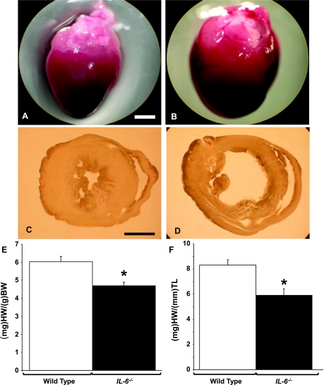
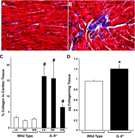
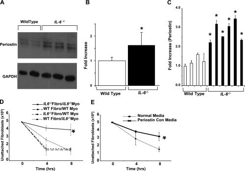
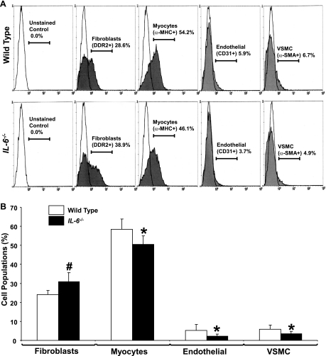
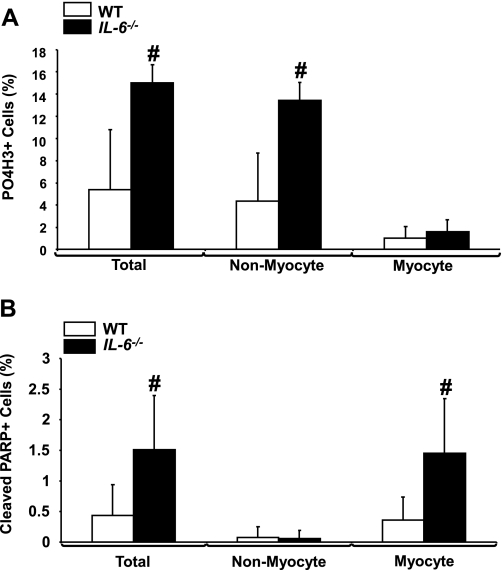
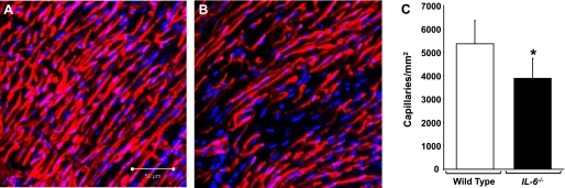
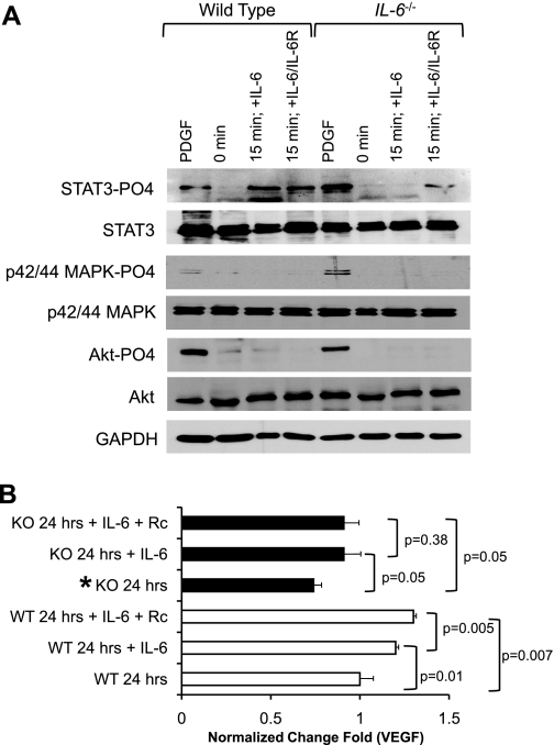
Interleukin-6 (IL-6) is a pleiotropic cytokine responsible for many different processes including the regulation of cell growth, apoptosis, differentiation, and survival in various cell types and organs, including the heart. Recent studies have indicated that IL-6 is a critical component in the cell-cell communication between myocytes and cardiac fibroblasts. In this study, we examined the effects of IL-6 deficiency on the cardiac cell populations, cardiac function, and interactions between the cells of the heart, specifically cardiac fibroblasts and myocytes. To examine the effects of IL-6 loss on cardiac function, we used the IL-6(-/-) mouse. IL-6 deficiency caused severe cardiac dilatation, increased accumulation of interstitial collagen, and altered expression of the adhesion protein periostin. In addition, flow cytometric analyses demonstrated dramatic alterations in the cardiac cell populations of IL-6(-/-) mice compared with wild-type littermates. We observed a marked increase in the cardiac fibroblast population in IL-6(-/-) mice, whereas a concomitant decrease was observed in the other cardiac cell populations examined. Moreover, we observed increased cell proliferation and apoptosis in the developing IL-6(-/-) heart. Additionally, we observed a significant decrease in the capillary density of IL-6(-/-) hearts. To elucidate the role of IL-6 in the interactions between cardiac fibroblasts and myocytes, we performed in vitro studies and demonstrated that IL-6 deficiency attenuated the activation of the STAT3 pathway and VEGF production. Taken together, these data demonstrate that a loss of IL-6 causes cardiac dysfunction by shifting the cardiac cell populations, altering the extracellular matrix, and disrupting critical cell-cell interactions.
Figures
References
-
- Ancey C, Menet E, Corbi P, Fredj S, Garcia M, Rücker-Martin C, Bescond J, Morel F, Wijdenes J, Lecron JC, Potreau D. Human cardiomyocyte hypertrophy induced in vitro by gp130 stimulation. Cardiovasc Res 59: 78–85, 2003. - PubMed
-
- Aukrust P, Ueland T, Lien E, Bendtzen K, Müller F, Andreassen AK, Nordøy I, Aass H, Espevik T, Simonsen S, Frøland SS, Gullestad L. Cytokine network in congestive heart failure secondary to ischemic or idiopathic dilated cardiomyopathy. Am J Cardiol 83: 376–382, 1999. - PubMed
-
- Banerjee I, Fuseler JW, Price RL, Borg TK, Baudino TA. Determination of cell type, and number during cardiac development in the neonatal and adult rat and mouse. Am J Physiol Heart Circ Physiol 293: H1883–H1891, 2007. - PubMed
-
- Banerjee I, Yekkala K, Borg TK, Baudino TA. Dynamic interactions between myocytes, fibroblasts, and extracellular matrix. Ann NY Acad Sci 1080: 76–84, 2006. - PubMed
Publication types
MeSH terms
Substances
Grants and funding
LinkOut - more resources
Full Text Sources
Molecular Biology Databases
Miscellaneous