. 2009 Mar 10;106(10):3794-9.
doi: 10.1073/pnas.0804543106. Epub 2009 Feb 20.

Endothelial expression of autocrine VEGF upon the uptake of tumor-derived microvesicles containing oncogenic EGFR

Affiliations

Endothelial expression of autocrine VEGF upon the uptake of tumor-derived microvesicles containing oncogenic EGFR

Khalid Al-Nedawi et al. Proc Natl Acad Sci U S A. .

Abstract

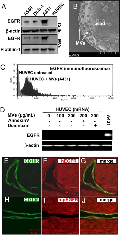
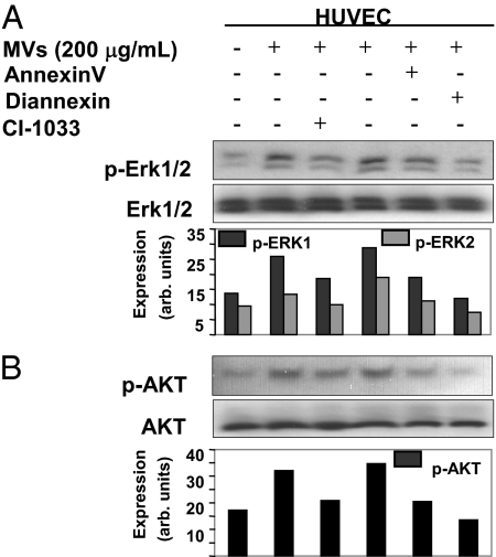
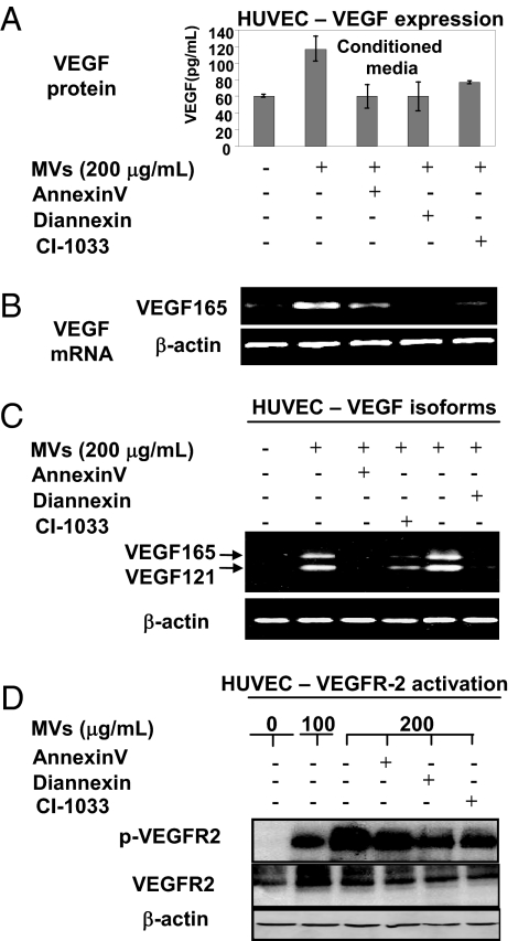
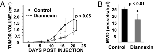
Activated EGF receptor (EGFR) plays an oncogenic role in several human malignancies. Although the intracellular effects of EGFR are well studied, its ability to induce and modulate tumor angiogenesis is less understood. We found previously that oncogenic EGFR can be shed from cancer cells as cargo of membrane microvesicles (MVs), which can interact with surfaces of other cells. Here we report that MVs produced by human cancer cells harboring activated EGFR (A431, A549, DLD-1) can be taken up by cultured endothelial cells, in which they elicit EGFR-dependent responses, including activation of MAPK and Akt pathways. These responses can be blocked by annexin V and its homodimer, Diannexin, both of which cloak phosphatidylserine residues on the surfaces of MVs. Interestingly, the intercellular EGFR transfer is also accompanied by the onset of VEGF expression in endothelial cells and by autocrine activation of its key signaling receptor (VEGF receptor-2). In A431 human tumor xenografts in mice, angiogenic endothelial cells stain positively for human EGFR and phospho-EGFR, while treatment with Diannexin leads to a reduction of tumor growth rate and microvascular density. Thus, we propose that oncogene-containing tumor cell-derived MVs could act as a unique form of angiogenesis-modulating stimuli and are capable of switching endothelial cells to act in an autocrine mode.


Conflict of interest statement

Conflict of interest statement: A.C.A. is employed by Alavita, which develops therapeutic applications of Diannexin, mainly in cardiovascular medicine.

Figures

Fig. 1.
Fig. 1.
Microvesicular transfer of the oncogenic EGFR to endothelial cells. (A) EGFR associated with cells and MVs. A549, DLD-1, and A431 cancer cells express high levels of EGFR and shed this oncogenic receptor into their conditioned medium as cargo of MVs. In contrast, neither HUVECs nor their derived MVs contain detectable EGFR. Protein loading was normalized to β-actin (cell lysates) or to flotillin-1 (MVs), a marker of membrane lipid rafts from which MVs originate (1, 8). (B) Scanning electron micrograph of A431-derived MVs adsorbed on poly-L-lysine beads. (Scale bar, 1 μm.) (C) Uptake of EGFR by HUVEC cells upon exposure to EGFR-containing MVs derived from cancer cells (A431). FACS analysis reveals a prominent shift in EGFR immunofluorescence of HUVECs after 24 h incubation with MVs followed by extensive washing. (D) Exposure to tumor cell-derived MVs does not induce endogenous EGFR gene expression in endothelial cells. HUVECs were treated with EGFR-containing MVs for 24 h and then processed for EGFR mRNA detection by RT-PCR. (E–G) Transfer of tumor-derived EGFR to endothelial cells in vivo. Co-localization of the human EGFR immunofluorescence (red) with staining for the mouse endothelial marker CD105 (green) in A431 xenografts (SCID mice). (H–J) Co-staining for phosphorylated human EGFR (h-pEGFR; red) and A431 tumor-associated mouse endothelium (CD105; green) (Scale bar, 10 μm.) For more information see Figs. S6–S8 and Movies S1–S3.
Fig. 2.
Fig. 2.
Signaling events triggered in endothelial cells upon the uptake of MV-derived EGFR. (A) Phosphorylation of MAPK (Erk1/2) in endothelial cells that have taken up EGFR upon incubation with MVs isolated from A431 conditioned medium. These effects were obliterated by preincubation of MVs with PS blocking agents: annexin V and Diannexin (both at 2 μg/mL) or with irreversible EGFR inhibitor CI-1033 (50 μM) before addition to HUVEC. The phospho-Erk1 (p-Erk1) signal was somewhat more robust than p-Erk2, and both were normalized to the corresponding Erk1/2 bands for the purpose of densitometric quantification, which is expressed in arbitrary units. (B) Impact of the microvesicular EGFR transfer on Akt phosphorylation in endothelial cells (HUVECs, all conditions as in A).
Fig. 3.
Fig. 3.
Microvesicular uptake of EGFR triggers VEGF expression and autocrine signaling in endothelial cells. (A) Secretion of VEGF into conditioned medium of serum-starved HUVECs upon their incubation with EGFR-containing MVs. This effect was markedly diminished by preincubation of MVs with agents blocking PS residues, annexin V and Diannexin (both at 2 μg/mL), and with the EGFR/panErb inhibitor CI-1033 (50 μM). The baseline level of VEGF represents the limit of detectability and should be considered as negative. (B) The onset of VEGF165 mRNA expression (RT-PCR) in HUVEC cells that have taken up MV-associated EGFR. Again, annexin V, Diannexin, and CI-1033 pre-treatments diminish the effects of MVs. (C) Expression of the main VEGF isoforms (VEGF165 and VEGF 121) in HUVECs is triggered by the transfer of tumor cell-derived MVs in a manner dependent on PS-mediated uptake and intact activity of the MV-associated EGFR. An independent set of primers was used to amplify VEGF121 and VEGF165 in HUVECs after exposure to either intact A431-derived MVs or MVs pretreated with annexin V, Diannexin, or CI-1033 (conditions as in A and B). (D) Autocrine activation of VEGFR-2 upon the uptake of EGFR-containing MVs by cultured endothelial cells (HUVECs). Serum starved HUVECs were treated with MVs as in A–C and assayed for VEGFR-2 phosphorylation (p-VEGFR-2). Interference with MV-related PS (annexin V and Diannexin) and EGFR kinase activity (i.e., CI-1033) diminishes the level of pVEGFR-2 in parallel with reduction of VEGF produced by HUVEC cells (compare A vs. C). The respective signals were quantified by densitometry, normalized to the total VEGFR-2, and expressed in relative units (Fig. S5).
Fig. 4.
Fig. 4.
Inhibition of tumor growth and angiogenesis by interference with membrane PS. (A) Delay of A431 tumor growth in SCID mice treated with daily doses of Diannexin (1 mg/kg i.p.). Average tumor volume ± SD; n = 5; P < 0.05. (B) Reduced MVD in A431 tumors exposed to Diannexin. Tumor sections were stained with anti-CD105 antibody to visualize endothelial cells and vascular counts were performed in “hot spots” at ×200 magnification.

References

    1. Al-Nedawi K, et al. Intercellular transfer of the oncogenic receptor EGFRvIII by microvesicles derived from tumour cells. Nat Cell Biol. 2008;10:619–624. - PubMed
    1. Pilzer D, Gasser O, Moskovich O, Schifferli JA, Fishelson Z. Emission of membrane vesicles: roles in complement resistance, immunity and cancer. Springer Semin Immunopathol. 2005;27:375–387. - PubMed
    1. Johnstone RM. Exosomes biological significance: a concise review. Blood Cells Mol Dis. 2006;36:315–321. - PubMed
    1. Janowska-Wieczorek A, et al. Microvesicles derived from activated platelets induce metastasis and angiogenesis in lung cancer. Int J Cancer. 2005;113:752–760. %20; - PubMed
    1. Mandal SK, Iakhiaev A, Pendurthi UR, Rao LV. Acute cholesterol depletion impairs functional expression of tissue factor in fibroblasts: modulation of tissue factor activity by membrane cholesterol. Blood. 2005;105:153–160. - PMC - PubMed

Publication types