. 2009 Mar 1;182(5):2628-40.
doi: 10.4049/jimmunol.0802954.

Transgenic inhibition of astroglial NF-kappa B improves functional outcome in experimental autoimmune encephalomyelitis by suppressing chronic central nervous system inflammation

Affiliations

Transgenic inhibition of astroglial NF-kappa B improves functional outcome in experimental autoimmune encephalomyelitis by suppressing chronic central nervous system inflammation

Roberta Brambilla et al. J Immunol. .

Abstract

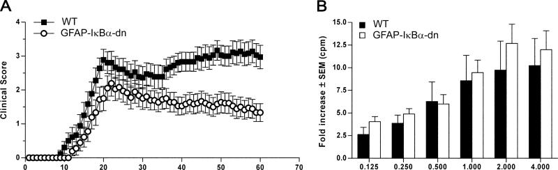
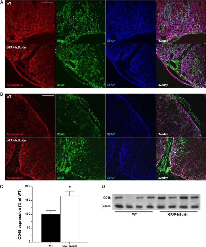
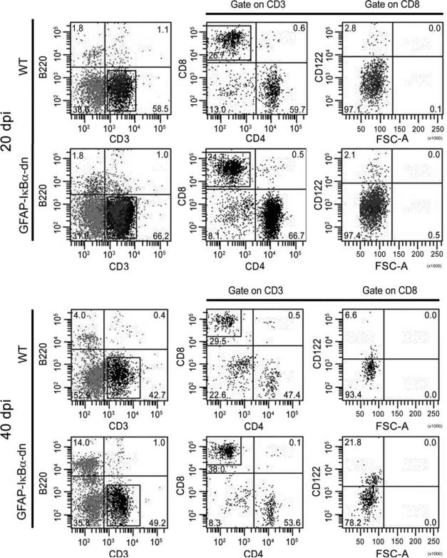
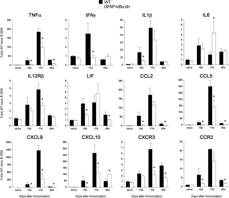
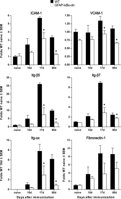
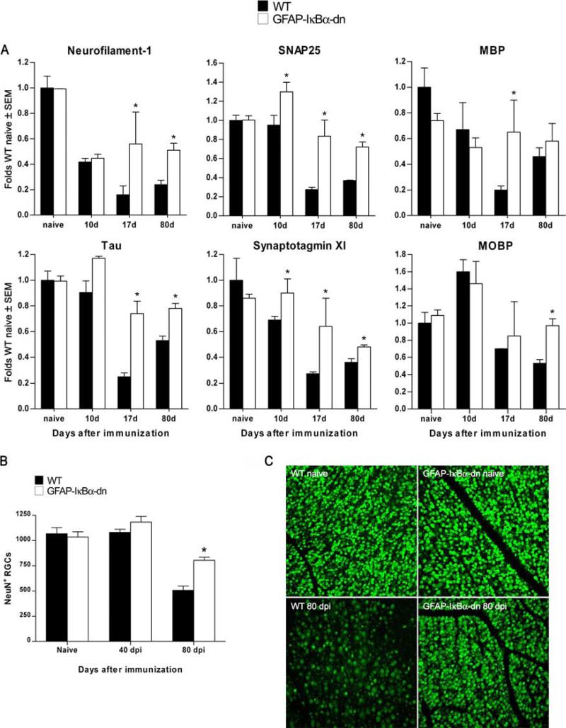
In the CNS, the transcription factor NF-kappaB is a key regulator of inflammation and secondary injury processes. Following trauma or disease, the expression of NF-kappaB-dependent genes is activated, leading to both protective and detrimental effects. In this study, we show that transgenic inactivation of astroglial NF-kappaB (glial fibrillary acidic protein-IkappaB alpha-dominant-negative mice) resulted in reduced disease severity and improved functional recovery following experimental autoimmune encephalomyelitis. At the chronic stage of the disease, transgenic mice exhibited an overall higher presence of leukocytes in spinal cord and brain, and a markedly higher percentage of CD8(+)CD122(+) T regulatory cells compared with wild type, which correlated with the timing of clinical recovery. We also observed that expression of proinflammatory genes in both spinal cord and cerebellum was delayed and reduced, whereas the loss of neuronal-specific molecules essential for synaptic transmission was limited compared with wild-type mice. Furthermore, death of retinal ganglion cells in affected retinas was almost abolished, suggesting the activation of neuroprotective mechanisms. Our data indicate that inhibiting NF-kappaB in astrocytes results in neuroprotective effects following experimental autoimmune encephalomyelitis, directly implicating astrocytes in the pathophysiology of this disease.


Figures

FIGURE 1
FIGURE 1
Inhibition of astroglial NF-κB improves functional outcome following EAE. A, Clinical course of MOG35–55-induced EAE in WT and GFAP-IκBα-dn mice. EAE symptoms were scored daily for 60 days, as described in Materials and Methods. Results are expressed as the daily mean clinical score ± SEM of 18 animals/group from two independent experiments. Curves are significantly different (p < 0.0001, Mann-Whitney U test). B, Cell proliferation assay on T cells isolated from WT and GFAP-IκBα-dn mice undergoing active EAE 2 wk after immunization. Following exposure to increasing MOG concentrations, cells were pulsed with [3H]thymidine, and proliferation was assessed in cpm of incorporated tritiated thymidine. Results are expressed as mean fold increase incorporation relative to background ± SEM of four samples/group.
FIGURE 2
FIGURE 2
Leukocyte infiltration in WT and GFAP-IκBα-dn spinal cords following EAE. A, Immunofluorescent labeling of WT and GFAP-IκBα-dn tissue sections at 20 dpi (acute disease). B, Immunofluorescent labeling of WT and GFAP-IκBα-dn tissue sections at 40 dpi (chronic disease). Red: Aquaporin-4 (localized to astrocyte end-feet in contact with the vascular endothelium). Green: pan-leukocyte marker CD45. Blue: astrocyte-specific marker GFAP. Scale bar: 100 μm. C, Quantification of CD45 protein expression in spinal cord tissue of WT and GFAP-IκBα-dn mice at 40 dpi. Data are normalized to β-actin. Results represent the mean ± SEM of six animals/group and are expressed as percentage of WT. *, p < 0.05; Student's t test. D, Representative Western blot probed for CD45 and β-actin as loading control.
FIGURE 3
FIGURE 3
Flow cytometric analysis of leukocyte infiltrates into the spinal cord of EAE-induced mice at 20 and 40 dpi. A representative experiment conducted by pooling eight animals/group is shown. The numbers in the quadrants represent the relative percentages of the various cell populations.
FIGURE 4
FIGURE 4
Differential expression of cytokines and chemokines in WT and GFAP-IκBα-dn spinal cord tissue following EAE. Gene expression was assessed in naive animals and at 10, 17, and 80 dpi. For each gene, results are expressed as folds of corresponding WT naive ± SEM after normalization to β-actin. Four animals/group/time were analyzed. *, p < 0.05 with respect to corresponding WT; one-way ANOVA, Tukey test.
FIGURE 5
FIGURE 5
TNF-α protein localization in WT and GFAP-IκBα-dn spinal cords following EAE. Immunofluorescent labeling of WT (A) and GFAP-IκBα-dn (B) tissue sections at 20 dpi (acute disease). Red: TNF-α. Green: GFAP or CD11b. Scale bar: 100 μm.
FIGURE 6
FIGURE 6
Differential expression of adhesion molecules and Itg in WT and GFAP-IκBα-dn mice following EAE. Gene expression was assessed in naive animals and at 10, 17, and 80 dpi. For each gene, results are expressed as folds of corresponding WT naive ± SEM after normalization to β-actin. Four animals/group/time were analyzed. *, p < 0.05 with respect to corresponding WT; one-way ANOVA, Tukey test.
FIGURE 7
FIGURE 7
Inhibition of astroglial NF-κB results in neuroprotective effects following EAE. A, Gene expression was assessed in naive animals and at 10, 17, and 80 dpi. For each gene, results are expressed as folds of corresponding WT naive ± SEM after normalization to β-actin. Four animals/group/time were analyzed. *, p < 0.05 with respect to corresponding WT; one-way ANOVA, Tukey test. B, Evaluation of RGC number on flat-mounted retinas labeled with FITC-conjugated NeuN Ab. C, Representative photomicrographs of flat-mounted retinas from WT and transgenic mice in naive and diseased (80 dpi) conditions. Nine animals per group were counted. *, p < 0.001 vs WT 80 days; one-way ANOVA, Tukey test. Scale bar = 100 μm.
FIGURE 8
FIGURE 8
Western blot quantification of CNS-specific proteins in WT and GFAP-IκBα-dn mice at 40 dpi. Quantification of GAP43 and MBP in spinal cord tissue of WT and GFAP-IκBα-dn mice at 40 dpi. Data are normalized to β-actin. Results represent the mean ± SEM of six animals/group and are expressed as percentage of WT. *, p < 0.05; Student's t test. Representative Western blots for each protein are shown.

References

    1. Kornek B, Storch MK, Weissert R, Wallstroem E, Stefferl A, Olsson T, Linington C, Schmidbauer M, Lassmann H. Multiple sclerosis and chronic autoimmune encephalomyelitis: a comparative quantitative study of axonal injury in active, inactive, and remyelinated lesions. Am. J. Pathol. 2000;157:267–276. - PMC - PubMed
    1. Ayers MM, Hazelwood LJ, Catmull DV, Wang D, McKormack Q, Bernard CC, Orian JM. Early glial responses in murine models of multiple sclerosis. Neurochem. Int. 2004;45:409–419. - PubMed
    1. Bjartmar C, Trapp BD. Axonal and neuronal degeneration in multiple sclerosis: mechanisms and functional consequences. Curr. Opin. Neurol. 2001;14:271–278. - PubMed
    1. Wujek JR, Bjartmar C, Richer E, Ransohoff RM, Yu M, Tuohy VK, Trapp BD. Axon loss in the spinal cord determines permanent neurological disability in an animal model of multiple sclerosis. J. Neuropathol. Exp. Neurol. 2002;61:23–32. - PubMed
    1. Babcock AA, Kuziel WA, Rivest S, Owens T. Chemokine expression by glial cells directs leukocytes to sites of axonal injury in the CNS. J. Neurosci. 2003;23:7922–7930. - PMC - PubMed

Publication types

MeSH terms