. 2009 May 1;284(18):12031-40.
doi: 10.1074/jbc.M808962200. Epub 2009 Feb 20.

Domain architecture of the stator complex of the A1A0-ATP synthase from Thermoplasma acidophilum

Affiliations

Domain architecture of the stator complex of the A1A0-ATP synthase from Thermoplasma acidophilum

Erik Kish-Trier et al. J Biol Chem. .

Abstract

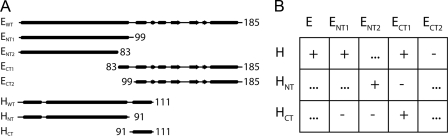
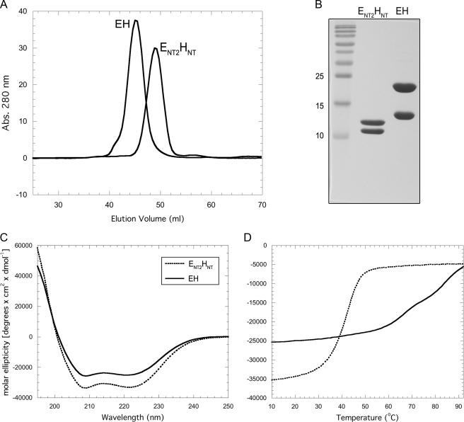
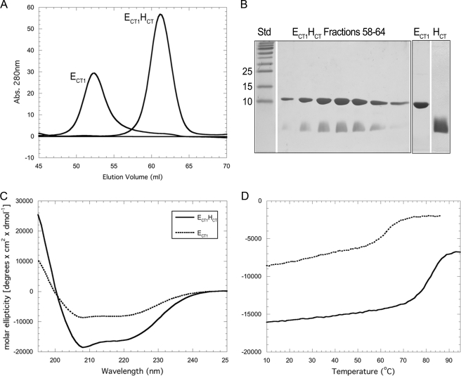
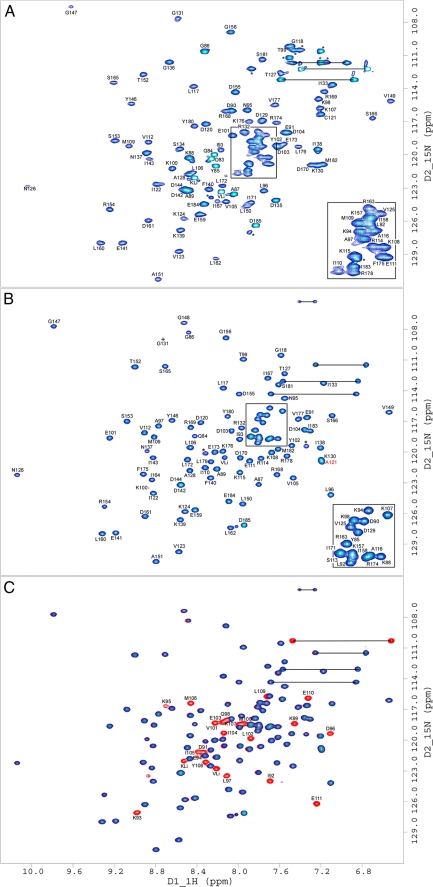
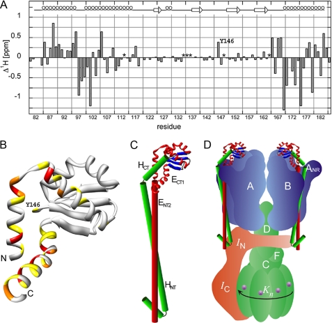
A key structural element in the ion translocating F-, A-, and V-ATPases is the peripheral stalk, an assembly of two polypeptides that provides a structural link between the ATPase and ion channel domains. Previously, we have characterized the peripheral stalk forming subunits E and H of the A-ATPase from Thermoplasma acidophilum and demonstrated that the two polypeptides interact to form a stable heterodimer with 1:1 stoichiometry (Kish-Trier, E., Briere, L. K., Dunn, S. D., and Wilkens, S. (2008) J. Mol. Biol. 375, 673-685). To define the domain architecture of the A-ATPase peripheral stalk, we have now generated truncated versions of the E and H subunits and analyzed their ability to bind each other. The data show that the N termini of the subunits form an alpha-helical coiled-coil, approximately 80 residues in length, whereas the C-terminal residues interact to form a globular domain containingalpha- and beta-structure. We find that the isolated C-terminal domain of the E subunit exists as a dimer in solution, consistent with a recent crystal structure of the related Pyrococcus horikoshii A-ATPase E subunit (Lokanath, N. K., Matsuura, Y., Kuroishi, C., Takahashi, N., and Kunishima, N. (2007) J. Mol. Biol. 366, 933-944). However, upon the addition of a peptide comprising the C-terminal 21 residues of the H subunit (or full-length H subunit), dimeric E subunit C-terminal domain dissociates to form a 1:1 heterodimer. NMR spectroscopy was used to show that H subunit C-terminal peptide binds to E subunit C-terminal domain via the terminal alpha-helices, with little involvement of the beta-sheet region. Based on these data, we propose a structural model of the A-ATPase peripheral stalk.


Figures

FIGURE 1.
FIGURE 1.
Summary of the EH truncation mutants and their interaction. A, schematic of full-length E and H subunits and truncation mutants used in this study. Secondary structure prediction was obtained from the PSIPRED prediction server. The arrows, cylinders, and lines denote predicted β-sheet, α-helix, and random coil content, respectively. B, table of interaction assay results. Positive and negative interactions are designated with (+ and -), indicating the ability/inability to co-purify by either ion exchange or gel filtration. Interactions not assessed in this study are marked (···).
FIGURE 2.
FIGURE 2.
Purification and characterization of ENT2HNT. A, comparison of EH and ENT2HNT gel filtration profiles. EH, which has been shown to be monodisperse and elongated in solution (12), elutes with an apparent molecular mass of ∼52 kDa (35.5 kDa actual). ENT2HNT elutes with a smaller apparent molecular mass of ∼43 kDa (21.5 kDa actual), consistent with ENT2HNT, like intact EH, being elongated and monodisperse. B, 15% SDS-PAGE gel of ENT2HNT and EH as visualized by Coomassie staining. Individual subunits were produced as fusion proteins and then reconstituted as described under “Experimental Procedures.” C, CD wavelength scan of ENT2HNT and EH in 10 mm sodium phosphate (pH 7) at 25 °C. Although both samples exhibit a union of the minima at 222 and 208 nm, indicating coiled-coil α-helical content, ENT2HNT exhibits ∼30% more molar ellipticity overall, suggesting that it represents the isolated coiled-coil domain. D, CD signal monitored at 222 nm as a function of increasing temperature (Tmelt). EH, as previously reported, exhibits several cooperative transitions, whereas ENT2HNT shows only a single strong transition, which may indicate it contains a single domain of EH, namely the coiled-coil domain. The reduced thermal stability may result from the isolation of the domain from the remainder of the complex.
FIGURE 3.
FIGURE 3.
Purification and characterization of ECT1 and ECT1HCT. A, overlay of ECT1 and ECT1HCT elution profiles collected during passage of the samples over a16 × 500 mm Superdex 75 gel filtration column. Binding of HCT to ECT1 resulted in an elution volume shift from 52 to 60.5 ml. This corresponds to a change in estimated molecular mass (spherical protein) from 35 to 22 kDa, compared with actual molecular masses of 12. kDa and 15 kDa for ECT1 and ECT1HCT, respectively. The change in elution volume suggests that HCT binding effectively reduced the spherical volume of ECT1, likely by out-competing homodimer formation. B, 15% SDS-PAGE gel of the ECT1HCT elution peak and individually purified components visualized by Coomassie staining. ECT1 and HCT were produced individually as fusion proteins and reconstituted as described under “Experimental Procedures.” C, CD wavelength scan of ECT1 and ECT1HCT in 10 mm sodium phosphate (pH 7) at 25 °C. Although retaining a shape indicative of mixed secondary structure, ECT1HCT exhibits a greater molar ellipticity than ECT1. Although this may be partially explained by the additional α-helical content contributed by HCT, it is also likely that HCT stabilizes ECT1 fold. This is further supported by the increase in thermal stability observed for ECT1HCT versus ECT1 alone (D) and additionally studied in Fig. 4.
FIGURE 4.
FIGURE 4.
15N HSQC NMR spectra of ECT1 and ECT1HCT. Two-dimensional 1H/15N HSQC NMR spectra taken at 298 K in 25 mm sodium phosphate (pH 7), 0.5 mm EDTA, and 0.02% NaN3. Spectra presented are: ECT1 (A), ECT1HCT where HCT is unlabeled for clarity (B), and where ECT1HCT is fully labeled (red) and is overlaid by the partially labeled spectrum from B in blue (C). The observed change in ECT1 behavior upon HCT binding, explored in Fig. 3, is further evidenced here by the global improvement in backbone amide peak signal dispersion and homogeneity indicative of ECT1 fold stabilization by HCT binding. Side chain amide peaks are linked by black horizontal lines. Resonances marked with an asterisk belong to amino acid side chains (e.g. Arg) or residues that could not be assigned because of overlap/poor signal to noise. KLi and VLi are part of the N-terminal linker (GPKVP). For ECT1 and ECT1HCT, Ser113, Gly148, Ile164, Ser134, Asp135, and Gly136 could not be assigned, respectively. The mutation Ta_E C121A has been described previously (Ref. ; indicated by the red label in B). The concentration of NMR samples was between 0.5 and 1 mm.
FIGURE 5.
FIGURE 5.
Secondary structure analysis of ECT1HCT as carried out by the chemical shift index protocol. A, the sequence alignment of the T. acidophilum ECT1 (Ta_ECT1) and the P. horikoshii ECT (Ph_ECT) was performed manually and refined by secondary structure comparison. Note that Ph_ECT has two α-helices connecting β-strands 1 and 2 that are significantly shorter in Ta_ECT1. The secondary structure for Ph_ECT (from the crystal structure; Ref. 11) and Ta_ECT1 (from the Hα and Cα chemical shift values using the chemical shift index (CSI) protocol as described under “Experimental Procedures”). The overall high degree of secondary structure similarity between the two proteins suggests that Ph_ECT is a good model for Ta_ECT1. Furthermore, the retention of secondary structure in Ta_ECT1 when bound to HCT, as shown here, indicates that the break up of Ta_ECT1 dimers by the addition of HCT (Figs. 3 and 4) is accompanied by a rearrangement of secondary structure elements rather than a change in composition of the same. B, the secondary structure analysis of HCT indicates α-helix for residues ∼95–108. See supplemental Fig. S4 for absolute deviations of 1Hα and 13Cα chemical shift values from random coil values.
FIGURE 6.
FIGURE 6.
Analysis of ECT1 chemical shift change upon the addition of HCT and a model of the proposed domain arrangement in the full-length EH peripheral stalk complex. Assignment of the 15N HSQC spectra presented in Fig. 4 permitted the calculation of ECT1 amide-proton chemical shift changes (Δ1Hδ) upon binding HCT. A, as can be seen, the majority of residues experiencing the most significant chemical shift perturbation upon HCT binding are located in the N- and C-terminal helices of ECT1 (residues for which no assignments could be obtained in either the ECT1 (Ser113, Gly148, and Ile164) or ECT1HCT (Ser134, Asp135, and Gly136) spectra are marked with an asterisk). B, the positions analogous to the Ta_ECT1 residues undergoing the largest chemical shift changes are highlighted in the available crystal structure of Ph_ECT with one dimer partner removed (Protein Data Bank code 2dma; Ref. 11). The largest change in chemical shift in the middle portion of the structure is experienced by Tyr146, which can be seen located close to the N- and C-terminal α-helices of Ph_ECT in the crystal structure (Ta_E Y146 corresponds to Ph_E M157). C, HCT, modeled as an α-helix and guided by the data presented here, is docked onto the Ph_ECT crystal structure monomer. Continuing below the globular ECT1HCT domain, the N-terminal domain, ENT2HNT, is modeled as a pair of parallel helices, representing the coiled-coil domain. We speculate that the N-terminal α-helix of HNT is folded back to interact with and stabilize the N-terminal ends of the coiled-coil domain. D, the resulting EH model placed into a schematic model of the T. acidophilum A-ATPase. The stoichiometry of the EH peripheral stalks has recently been determined for the related A/V-ATPase from T. thermophilus (6).

References

    1. Müller, V., and Grüber, G. (2003) Cell. Mol. Life. Sci. 60 474-494 - PMC - PubMed
    1. Forgac, M. (2007) Nat. Rev. Mol. Cell Biol. 8 917-929 - PubMed
    1. Boyer, P. D. (1997) Annu. Rev. Biochem. 66 717-749 - PubMed
    1. Wilkens, S. (2005) Adv. Prot. Chem. 71 345-382 - PubMed
    1. Coskun, U., Chaban, Y. L., Lingl, A., Müller, V., Keegstra, W., Boekema, E. J., and Grüber, G. (2004) J. Biol. Chem. 279 38644-38648 - PubMed

Publication types

MeSH terms

LinkOut - more resources