. 2009 Mar 12;458(7235):172-7.
doi: 10.1038/nature07836. Epub 2009 Feb 22.

Membrane scission by the ESCRT-III complex

Affiliations

Membrane scission by the ESCRT-III complex

Thomas Wollert et al. Nature. .

Abstract

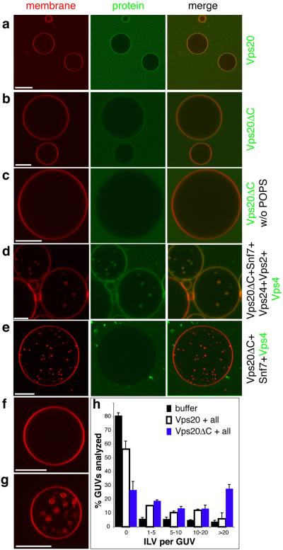
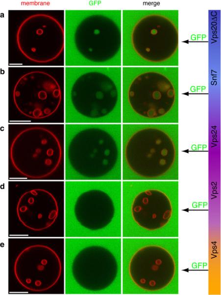
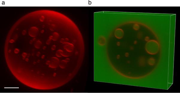
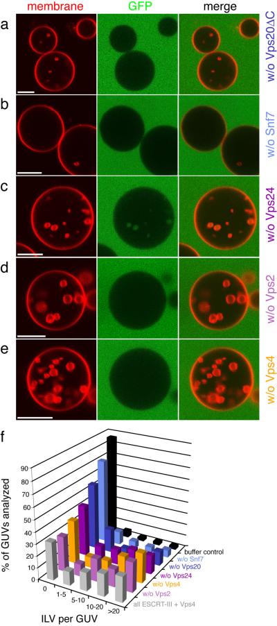
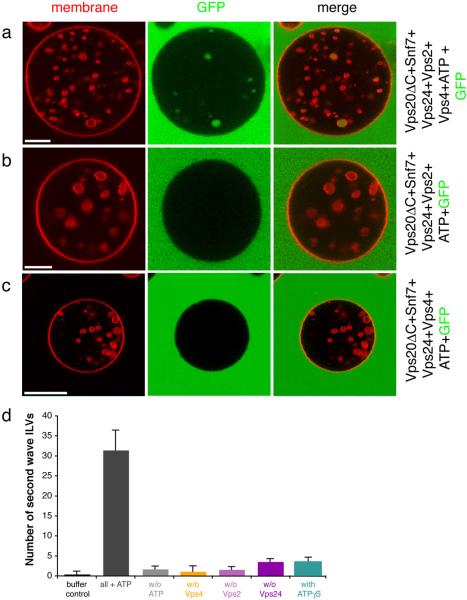
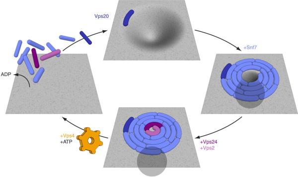
The endosomal sorting complex required for transport (ESCRT) system is essential for multivesicular body biogenesis, in which cargo sorting is coupled to the invagination and scission of intralumenal vesicles. The ESCRTs are also needed for budding of enveloped viruses including human immunodeficiency virus 1, and for membrane abscission in cytokinesis. In Saccharomyces cerevisiae, ESCRT-III consists of Vps20, Snf7, Vps24 and Vps2 (also known as Did4), which assemble in that order and require the ATPase Vps4 for their disassembly. In this study, the ESCRT-III-dependent budding and scission of intralumenal vesicles into giant unilamellar vesicles was reconstituted and visualized by fluorescence microscopy. Here we show that three subunits of ESCRT-III, Vps20, Snf7 and Vps24, are sufficient to detach intralumenal vesicles. Vps2, the ESCRT-III subunit responsible for recruiting Vps4, and the ATPase activity of Vps4 were required for ESCRT-III recycling and supported additional rounds of budding. The minimum set of ESCRT-III and Vps4 proteins capable of multiple cycles of vesicle detachment corresponds to the ancient set of ESCRT proteins conserved from archaea to animals.


Figures

Fig. 1
Fig. 1
ESCRT-III and Vps4 bind to GUV membranes. The GUV membrane is stained red with rhodamine-PE, and selected proteins are labeled green with Alexa-488. a, Vps20 and b, Vps20ΔC are recruited to membranes containing 10% POPS and 3% PI(3)P. c, Vps20ΔC is not recruited to membranes lacking POPS. d, Vps4 colocalizes with GUVs in the presence of Vps20ΔC, Snf7, Vps2, and Vps24. e, Vps4 is not recruited to GUVs if Vps2 and Vps24 are omitted. f, Five rounds of buffer addition to GUVs leads to no ILV formation. g, Vps20ΔC, Snf7, Vps24, Vps2 and Vps4 generate ILVs. h, ILVs were counted in a total 100 GUVs for each condition shown. Error bars were calculated using two independent experiments and calculating the difference between them. “All” refers to Snf7, Vps24, Vps2 and Vps4. Scale bar = 10 μm.
Fig. 2
Fig. 2
Uptake of the bulk phase and vesicle scission by ESCRT-III. GUVs (red) were incubated with all of Vps20ΔC, Snf7, Vps24, Vps2 and Vps4 in each experiment shown. Each component was added sequentially and incubated 5 min before the addition of the next component. To determine whether ILVs are connected with the bulk phase, the soluble marker GFP (green) was added immediately after either Vps20 ΔC (a), Snf7 (b), Vps24 (c), Vps2 (d), or Vps4 (e). No ATP or other nucleotide was present at any time. These data show that until Vps2 was added, the ILVs were connected with the bulk phase. Scale bar = 10 μm.
Fig. 3
Fig. 3
3-D reconstruction of an ESCRT-III treated GUV. a, Three-dimensional reconstruction of a GUV that was incubated with Vps20ΔC, Snf7, Vps24, Vps2, and Vps4, with GFP added together with Snf7. b, A Z stack of the same GUV shows that the ILVs are filled with GFP indicating that they were detached subsequent to the addition of Snf7. Scale bar = 5 μm.
Fig. 4
Fig. 4
Contributions of individual ESCRT-III subunits to ILV formation. GUVs were incubated with Vps20ΔC, Snf7, Vps24, Vps2 and Vps4, except that one component at a time was omitted. Omission of Vps20ΔC (a) or Snf7 (b) reduces ILV formation dramatically. Omitting Vps24 (c) leads to delayed detachment of ILVs whereas omission of Vps2 (d) and Vps4 (e) has no significant effect. f, Summary of the omission experiment (n = 100). Each color corresponds to a single experiment where the indicated component was omitted, buffer control - only buffer has been added to GUVs (black). Grey - positive control, reaction initiated in the presence of Vps20 ΔC, Snf7, Vps24, Vps2, and Vps4. Scale bar = 10 μm.
Fig. 5
Fig. 5
Vps4 and ATP induce a second cycle of ILV formation. a, GUVs (red) were incubated with Vps20ΔC, Snf7, Vps24, Vps2, and Vps4. GFP (green) was added after Vps4. In the absence of Vps4 (b) or Vps2 (c), no GFP-filled ILVs are formed following ATP addition. Scale bar = 10 μm. d, Quantification of the ATP induced second wave of ILV formation. Buffer control: only buffer and ATP were added to GUVs (black bar). Grey bars: GUVs with all protein components (Vps20ΔC, Snf7, Vps24, Vps2, and Vps4) in the presence (dark grey) and absence (light grey) of ATP. Colored bars: the indicated subunit was omitted from the reaction in the presence of all other components and ATP. Two sets of 15 GUVs were analyzed for each reaction and the difference between the two sets was used to calculate error bars.
Fig. 6
Fig. 6
A simple hypothesis for the mechanism of the ESCRT-III-Vps4 membrane scission cycle. The spiral structure is inspired by the observations of Hanson, Heuser, and colleagues . ESCRT-III sequesters cargo in MVBs, with Snf7 playing the most important role of the four subunits . Any cargo present in the neck near the growing array would be forced out of the neck and further into the bud at this stage, leading to its sequestration. Completion of the spiral would bring the membrane lipids of the neck into critical proximity, allowing fusion. The fused neck would then resolve into the detached vesicle on the interior and the ESCRT-III-coated limiting membrane on the exterior (bottom panel).

Comment in

References

    1. Gruenberg J, Stenmark H. The biogenesis of multivesicular endosomes. Nat. Rev. Mol. Cell Biol. 2004;5:317–323. - PubMed
    1. Russell MRG, Nickerson DP, Odorizzi G. Molecular mechanisms of late endosome morphology, identity and sorting. Curr. Opin. Cell Biol. 2006;18:422–428. - PubMed
    1. Slagsvold T, Pattni K, Malerod L, Stenmark H. Endosomal and nonendosomal functions of ESCRT proteins. Trends Cell Biol. 2006;16:317–326. - PubMed
    1. Saksena S, Sun J, Chu T, Emr SD. ESCRTing proteins in the endocytic pathway. Trends Biochem. Sci. 2007;32:561–573. - PubMed
    1. Piper RC, Katzmann DJ. Biogenesis and function of multivesicular bodies. Annu. Rev. Cell Devel. Biol. 2007;23:519–547. - PMC - PubMed

Publication types

MeSH terms

Substances