. 2009 Mar;11(3):286-94.
doi: 10.1038/ncb1836. Epub 2009 Feb 22.

Electrochemical cues regulate assembly of the Frizzled/Dishevelled complex at the plasma membrane during planar epithelial polarization

Affiliations

Electrochemical cues regulate assembly of the Frizzled/Dishevelled complex at the plasma membrane during planar epithelial polarization

Matias Simons et al. Nat Cell Biol. 2009 Mar.

Erratum in

  • Nat Cell Biol. 2009 Apr;11(4):508

Abstract

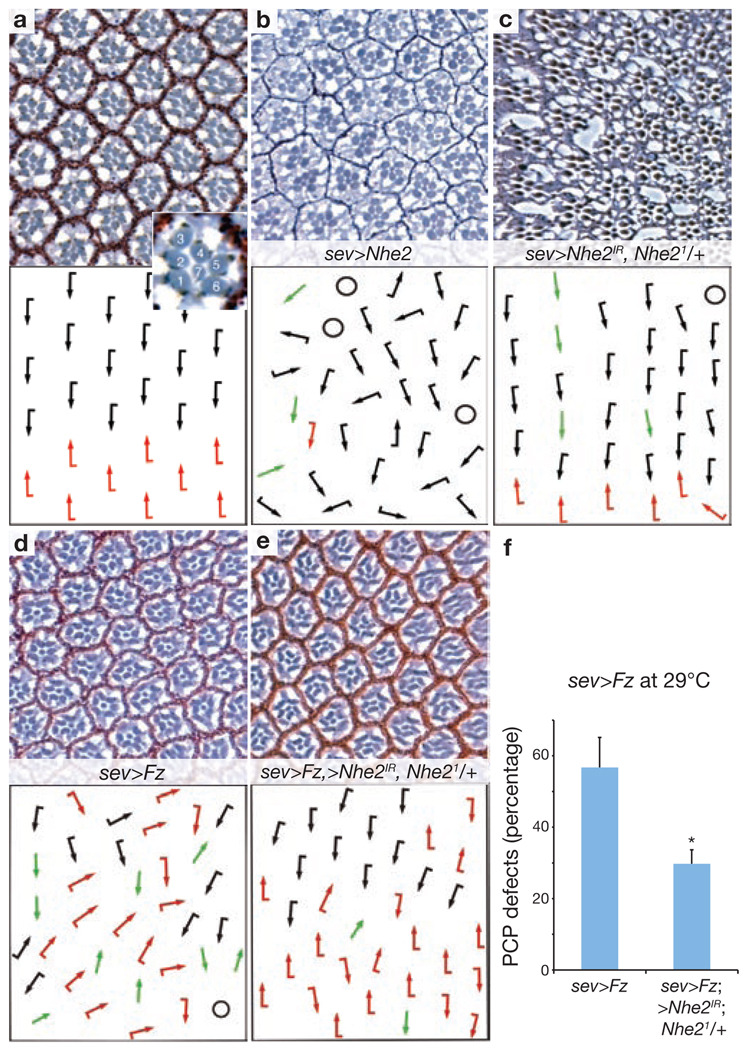
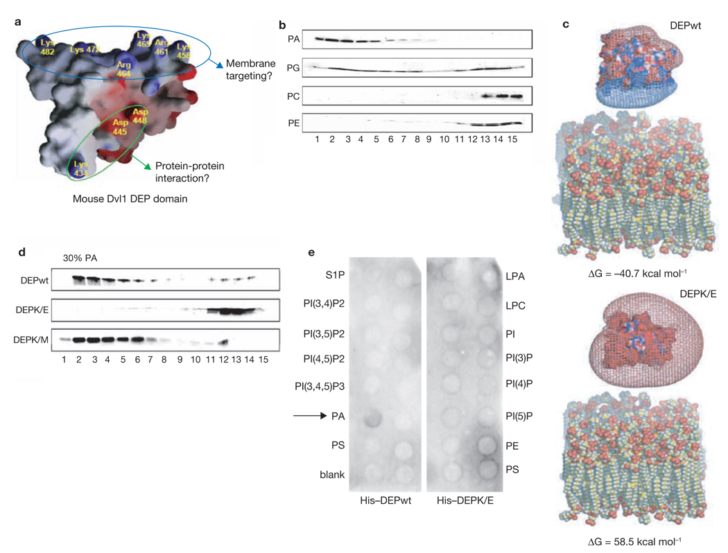
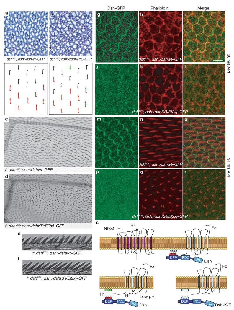
Dishevelled (Dsh) is a cytoplasmic multidomain protein that is required for all known branches of the Wnt signalling pathway. The Frizzled/planar cell polarity (Fz/PCP) signalling branch requires an asymmetric cortical localization of Dsh, but this process remains poorly understood. Using a genome-wide RNA interference (RNAi) screen in Drosophila melanogaster cells, we show that Dsh membrane localization is dependent on the Na(+)/H(+) exchange activity of the plasma membrane exchanger Nhe2. Manipulating Nhe2 expression levels in the eye causes PCP defects, and Nhe2 interacts genetically with Fz. Our data show that the binding and surface recruitment of Dsh by Fz is pH- and charge-dependent. We identify a polybasic stretch within the Dsh DEP domain that binds to negatively charged phospholipids and appears to be mechanistically important. Dsh recruitment by Fz can be abolished by converting these basic amino-acid residues into acidic ones, as in the mutant, DshKR/E. In vivo, the DshKR/E(2x) mutant with two substituted residues fails to associate with the membrane during active PCP signalling but rescues canonical Wnt signalling defects in a dsh-background. These results suggest that direct interaction between Fz and Dsh is stabilized by a pH and charge-dependent interaction of the DEP domain with phospholipids. This stabilization is particularly important for the PCP signalling branch and, thus, promotes specific pathway selection in Wnt signalling.


Figures

Figure 1
Figure 1
Nhe2/NHE3 activity is required for Fz-mediated Dsh recruitment. (a) Genome-wide RNAi library screen for Fz-mediated Dsh recruitment in D. melanogaster S2R+ cells. The cells were plated in 57 384-well plates containing dsRNA against 90% of D. melanogaster genes, transfected with myc–fz and dsh–GFP plasmids and analysed visually (see Methods). (b, c) Screen hits were defined by a × 20 microscope field with >10 cells showing defective Dsh–GFP recruitment. Control (Relish dsRNA) showed complete recruitment (b, see enlarged cell, inset), Nhe2 dsRNA caused defects in recruitment (c), see arrowheads and inset for defects. (d, e) HEK293T cells expressing Myc–Fz and Dsh–GFP were treated with the NHE3 inhibitor S3226 at 50 µM for 12 h. Treatment led to a redistribution of Dsh–GFP (e) as opposed to the control (DMSO) (d). (f) NHE3 knockdown caused defective Dsh recruitment (64.3% ± 2.3%, compared with 31.6% ± 4.3% in controls, this baseline defect in Dsh recruitment is due to heterogenous Fz and Dsh expression in these cells). Treatment with 30 mM NH4Cl to increase pHi (mean ± s.e.m. from three independent experiments, measured in f, right graph) rescued the recruitment defect in NHE3-knockdown cells (mean ± s.d., n= three independent experiments, *P < 0.001, measured in f left graph). (g–i) Alkalinization was transient with gradual pH normalization within 20 min. Images for all three conditions in f are shown. (j–p) Intracellular acidification interferes with Dsh recruitment. S2R+ cells were prepulsed for 30 min with 30 mM NH4Cl and then switched to a Na+-free medium for 12–16 h. This treatment caused a stable reduction of pHi to 7.1 (measured by E2GFP expression and pH calibration), resulting in defective Dsh recruitment (k–m). Incubation in a Na+-free medium without prepulsing (pHi 7.28) or in Na+-containing medium after prepulsing (mean ± s.d., n = 4 independent experiments, *P < 0.001) mildly affected recruitment. For quantification (j), only cells expressing Fz at their cell surface were counted. Note robust recruitment in untreated cells with pHi 7.45. (n–p) In a second assay, pHi was lowered by applying K+/nigericin for 4 h and clamping pHi according to an extracellular buffer (pH 6.8). This did not affect membrane association of the phosphatidylserine-binding LactC2–RFP but did affect Dsh membrane localization. Scale bars represent 10 µm.
Figure 2
Figure 2
Nhe2 shows gain- and loss-of-function PCP phenotypes in D. melanogaster eye, and interacts genetically with Fz. (a–e) show tangential eye sections of adult eyes (upper panels) with respective schematic representations (lower panels). Sections are around the equator (except in b, which shows a dorsal area). Wild-type eye with ommatidia of opposing chirality arranged around the equator and R3 positioned at the tip of the trapezoid is shown in a. Dorsal and ventral ommatidia are depicted with black and red arrows, respectively. Inset shows enlarged ommatidium with numbered photoreceptor cells. Overexpression of the UAS-Nhe2short splice variant (and also the long form, data not shown) with sev>GAL4 caused PCP defects (b) consisting of symmetrical clusters (green arrows), rotation defects and occasional chirality inversions. Extra photoreceptors, as well as fused ommatidia, were also seen. Circles represent unscorable clusters. Knockdown of Nhe2 and removal of one copy of Nhe2 (sev>Nhe2IR; Nhe21/+) led to PCP defects (c, 7.11%±3.5%, data are mean ± s.d of three independent eyes with > 400 ommatidia scored). In addition, general defects of eye morphology and structure are commonly seen with this genotype. (d–f) The sev>Fz phenotype is suppressed by co-expression of UAS-Nhe2IR and removal of one Nhe2 copy. This experiment was performed at 29 °C to enhance Fz and Nhe2IR expression. Total PCP defects were scored and quantified (f , data are mean ± s.d. of six eyes with > 700 ommatidia scored, *P < 0.0001).
Figure 3
Figure 3
The Dsh DEP domain interacts with acidic phospholipids. (a) The solution structure of the DEP domain of mouse Dvl1 shows a polybasic amino acid stretch positioned at a different surface location than the electric dipole for putative protein–protein interaction (which includes the lysine mutated in dsh1-K/M). (b) SUV liposomes consisting of 50% PC and 50% of the indicated phospholipids were mixed with the Dvl1 DEP domain and separated by chromatography on a Sephacryl S-300 column. The column fractions were analyzed by silver-staining. The lipid-bound DEP domain was eluted from fractions 1 to 7, whereas free DEP domain was eluted from fractions 10 to 15. (c) Electrostatic potentials were calculated using the non-linear Poisson-Boltzmann equation, to model phospholipid bilayers containing a 1:1 mixture of PC and PA. The complexity of the structures of DEPwt, DEPK/E and the lipid membrane model was minimized using a molecular dynamic simulation method (see Methods). The binding energy (ΔG) between DEPwt and the acidic lipid membrane model was predicted to be −40.7 kcal mol−1 (upper panel). However, for the DEPK/E mutant an unfavorable binding energy of 58.5 kcal mol−1 was calculated (lower panel). (d) SUV liposomes containing 70% PC and 30% PA were mixed with DEPwt, DEPK/E and DEPK/M (Lys 434, see a for location of mutations); binding to DEPK/E was markedly decreased. The overall structure of the isolated Dvl1 DEP domain was not affected by the K/E mutations as determined by chemical shift patterns from NMR spectroscopy (data not shown). Full scans of b and d are shown in Supplementary Information, Fig. S6. (e) Nitrocellulose filters (PIP strips) spotted with several different phospholipids species were overlayed with His-tagged DEPwt and DEPK/E (1 µg ml−1) and subjected to western blotting with a monoclonal anti-His antibody. DEPwt bound specifically to PA (arrow). DEPK/E did not bind to any of the spotted phospholipids. LPA, lysophosphatidic acid; LPC, lysophosphatidylcholine; PI, phosphatidylinositol; PIXPn, phosphatidylinositol X phosphaten; PE, phosphatidylethanolamine; PC, phosphatidylcholine; S1P, sphingosine-1-phosphate; PA, phosphatidic acid; PS, phosphatidylserine.
Figure 4
Figure 4
Fz-mediated Dsh membrane recruitment is charge-dependent. (a–f) DshKR/E(5×)–GFP, bearing five lysine or arginine substitutions for glutamic acid, was no longer recruited by Fz in S2R+ cells (d–f). The mutated amino acids correspond to the conserved residues mutated in mouse Dvl1 DEP-K/E. Fz efficiently recruited Dshwt (ac). Fz was visualized with a monoclonal anti-Myc antibody. Dsh-(KR/E) is expressed in higher amounts than Dshwt, as shown by western blotting (Supplementary Information, Fig. S5b). Merged panels (c, f) also include Hoechst 33342 (blue) as nuclear stain. (g–l) DshKR/E–GFP shows defective recruitment to the apical junctions by Myc–Fz in third-instar wing discs in vivo. Dshwt–GFP (g) and DshKR/E–GFP (j) were expressed ubiquitously under endogenous dsh promoter sequences. Myc–Fz (h, k) was expressed in a stripe at the anterior-posterior boundary using dpp>GAL4. Displayed is a x/z-stack of wing epithelial cells with apical at the top and basal at the bottom. Unlike Dshwt–GFP (arrows, g–i), DshKR/E–GFP is not recruited to the apical membrane by Fz (arrowheads, j–l). Other than the dpp stripe, the expression strength and localization patterns of Dshwt and DshKR/E(5×) are indistinguishable (g, j). (m–r) Neutralization of negatively charged membrane lipids (such as PA) by the membrane-permeant weak base sphingosine (75 µM), affects Dsh–GFP localization in HEK293T cells. The overlap of Dsh and Fz at the plasma membrane is diminished (p–r), compared with control vehicle-treated (ethanol) cells (m–o). Partial internalization of both proteins is also seen. The same pattern is observed in S2R+ cells, data not shown. (s–u) Sphingosine rescues the defective recruitment of DshKR/E–GFP by Fz. Sphingosine (75 µM) and ethanol were applied for 3 h. Sphingosine rescue was quantified (u), data are mean ± s.d, n = 10 different microscope fields from two experiments, *P < 0.0001. (v–x) DshKR/E–GFP co-localizes with Fz in sphingosine-rescued cells.
Figure 5
Figure 5
The polybasic stretch in the Dsh DEP domain is essential for PCP signalling in vivo. Dshwt–GFP and DshKR/E–GFP were expressed under a dsh promoter in a dshV26 null background (dshV26; dsh>dsh–GFP). (a–r) Flies that rescued the dshV26 lethality were scored for phenotypes in adult eyes (a, b, schematic representations lower panels, dorsal and ventral ommatidia are depicted with black and red arrows respectively, the green arrow depicts a symmetrical cluster), adult wings (c–f) and pupal wings (g–r). In the eye, DshKR/E(2×) caused mild phenotypes, including symmetrical clusters, chirality inversions and rotation defects (b). In adult wings, DshKR/E(2×) caused severe defects in wing-hair orientation, whereas the complete wing margin was intact (d). The anterior wing margin bristles are shown for DshKR/E(2×) (f), and Dshwt (e). Rescue with Dshwt was complete with fully wild-type appearance (a, c, e). (g–r) Pupal wings were examined at a stage before wing-hair formation (~30 h APF, g–l) and during wing hair formation (~34 hours APF, m–r). Dshwt–GFP co-localized with phalloidin-stained actin at the cell cortex at the early stage (g–i). In contrast, DshKR/E(2×)–GFP showed diffuse cytoplasmic localization (j–l). At the later stage (~34 h APF), Dshwt–GFP showed a typical cortical distribution with enrichment in the proximo-distal axis, and wing hairs showed a normal proximo-distal orientation (m–o, as reflected by Phalloidin, red). DshKR/E(2×)–GFP also failed at this stage to associate with the membrane (p–r). The resulting phenotypes are misoriented wing hairs and a multiple wing hair phenotype (q–r). Wing hairs appear shorter than shown in n, as the misoriented hairs point up and out of the confocal plane. (s) Suggested model of Fz-mediated Dsh membrane recruitment. Dsh binds weakly via its PDZ domain to the Fz C terminus. In addition, the DEP domain binds directly to acidic phospholipids. The interaction is dependent on local pH and charge. With Dshwt , proximity of the Na+/H+ exchanger Nhe2 to Fz maintains a slightly basic local pH (upper panel). When local pH is lower (Nhe2 mutant), the lipid headgroups are protonated, losing their negative charge (green) and leading to repulsion of Dsh (lower left panel). Similarly, mutations in the polybasic stretch of the DEP domain cause cytoplasmic Dsh localization (lower right panel), which can be rescued by lowering surface negativity (Fig. 4s–u). It is possible that another surface of the DEP domain also interacts with Fz directly.

Comment in

References

    1. Boutros M, Paricio N, Strutt DI, Mlodzik M. Dishevelled activates JNK and discriminates between JNK pathways in planar polarity and wingless signaling. Cell. 1998;94:109–118. - PubMed
    1. Seifert JR, Mlodzik M. Frizzled/PCP signalling: a conserved mechanism regulating cell polarity and directed motility. Nature Rev. Genet. 2007;8:126–138. - PubMed
    1. Veeman MT, Axelrod JD, Moon RT. A second canon. Functions and mechanisms of β-catenin-independent Wnt signaling. Dev. Cell. 2003;5:367–377. - PubMed
    1. Axelrod JD. Unipolar membrane association of Dishevelled mediates Frizzled planar cell polarity signaling. Genes Dev. 2001;15:1182–1187. - PMC - PubMed
    1. Bilic J, et al. Wnt induces LRP6 signalosomes and promotes dishevelled-dependent LRP6 phosphorylation. Science. 2007;316:1619–1622. - PubMed

Publication types

MeSH terms