. 2009 Mar;15(3):277-84.
doi: 10.1038/nm.1929. Epub 2009 Feb 22.

Effector T cells control lung inflammation during acute influenza virus infection by producing IL-10

Affiliations

Effector T cells control lung inflammation during acute influenza virus infection by producing IL-10

Jie Sun et al. Nat Med. 2009 Mar.

Abstract

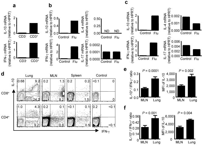
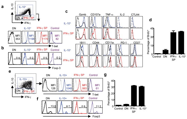
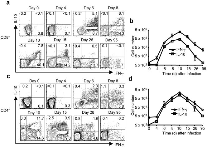
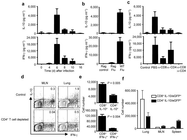
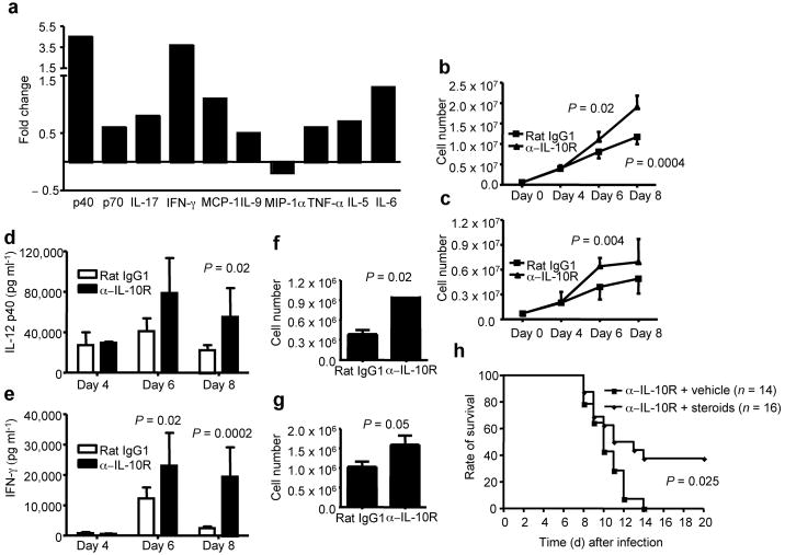
Activated antigen-specific T cells produce a variety of effector molecules for clearing infection but also contribute to inflammation and tissue injury. Here we report an anti-inflammatory property of antiviral CD8+ and CD4+ effector T cells (T(eff) cells) in the infected periphery during acute virus infection. We find that, during acute influenza infection, interleukin-10 (IL-10) is produced in the infected lungs in large amounts--exclusively by infiltrating virus-specific T(eff) cells, with CD8+ T(eff) cells contributing a larger fraction of the IL-10 produced. These T(eff) cells in the periphery simultaneously produce IL-10 and proinflammatory cytokines and express lineage markers characteristic of conventional T helper type 1 or T cytotoxic type 1 cells. Notably, blocking the action of the T(eff) cell-derived IL-10 results in enhanced pulmonary inflammation and lethal injury. Our results show that antiviral T(eff) cells exert regulatory functions--that is, they fine-tune the extent of lung inflammation and injury associated with influenza infection by producing an anti-inflammatory cytokine. We discuss the potential implications of these findings for infection with highly pathogenic influenza viruses.


Conflict of interest statement

COMPETING INTERESTS STATEMENT

The authors declare that they have no competing financial interests.

Figures

Figure 1
Figure 1. Influenza-specific T cells preferentially produce IL-10 and IFN-γ in the influenza-infected BALB/c lungs
(a) Quantitative RT-RCR analysis of IL-10 and IFN-γ transcript levels of purified CD3+and CD3 cells in the lungs of mice 8 days p.i. (b, c) Quantitative RT-RCR analysis of IL-4, IL-17, IL-10 and IFN-γ transcript levels of purified CD8+(b) or CD4+(c) T cells in the lungs of mice 8 days p.i. (d) At day 8 p.i., lung, mediastinal lymph node (MLN) and spleen cells were restimulated in vitro with influenza infected BMDC and intracellular cytokine staining (ICS) was performed to measure the production of IL-10 and IFN-γ in gated CD8+ and CD4+ T cells. Numbers indicate the percentages of cytokine-positive cells in the gates within the total population. (e, f) Cells from MLN and lungs of mice 8 days p.i. were restimulated with flu-infected BMDC and ICS was performed to measure the production of IL-10 and IFN-γ in gated CD8+ (e) and CD4+ (f) T cells. The ratio of IL-10+ vs. IFN-γ+ CD8+ (e) or CD4+ (f) T cells and the mean fluorescence intensity (MFI) of IL-10 staining in IL-10+ CD8+ (e) or IL-10+ CD4+ (f) T cells in MLN and lungs are depicted. P values were determined by unpaired two-tailed student t test.
Figure 2
Figure 2. IL-10 producing CD8+ and CD4+ T cells are Type 1 effectors
BALB/c mice were infected with influenza. CD8+ (a–c) or CD4+ (e, f) cells from lungs of uninfected mice or mice 7 days p.i. with influenza were restimulated with influenza infected BMDC and the expression of T-bet (a, e), Foxp-3 (b, f) and a panel of effector or activation molecules (c) in uninfected lung CD8+ or CD4+ T cells (control) or IL-10 positive CD8+ or CD4+ T cells (IL-10+), IFN-γ single positive CD8+ or CD4+ T cells (IFN-γ SP), IL-10 and IFN-γ double negative CD8+ or CD4+ T cells (DN) from infected lungs were measured by ICS. Gray line: isotype control Ab staining. Numbers are MFI (a, e) or percentages of Foxp-3-positive cells in the gates within the total population (b, f). (d, g) Uninfected mice (control) and mice 6 days p.i. were injected with BrdU. 2 h later, mice were sacrificed and lung cells were restimulated with influenza-infected BMDC. The percentages of BrdU+ cells in CD8+ (d) or CD4+ (g) cells of uninfected lung (control) and the IL-10+, IFN-γ single positive (IFN-γ SP) and IL-10 IFN-γ (DN) T cells of 6 days p.i. lung are depicted.
Figure 3
Figure 3. Kinetics of IL-10 producing CD8+ and CD4+ Te in vivo.
BALB/c mice were infected with influenza. At the indicated days p.i, lung cells were restimulated with influenza-infected BMDC. The production of IL-10 and IFN-γ by CD8+ and CD4+ T cells was measured by ICS. (a, c) FACS plots of IL-10 and IFN-γ production by CD8+ (a) or CD4+ (c) T cells in the lung Numbers indicate the percentages of cytokine-positive cells in the gates within the CD8+ (a) or CD4+ (c) T cell population. (b, d) The kinetics of accumulation (absolute numbers) of IL-10+ or IFN-γ+ CD8+ T cells (b) or IL-10+ or IFN-γ+ CD4+ T cells (d) in the lungs following influenza infection.
Figure 4
Figure 4. Te are the major source of IL-10 in vivo during influenza infection
BALB/c mice were infected with influenza (a–e). (a) At the indicated days p.i., IL-10 and IFN-γ levels in the BALF were measured by ELISA. (b) WT and Rag1−/− mice were infected with influenza. At day 6 p.i., IL-10 and IFN-γ levels in the BALF were measured by ELISA. Rag control (Rag1−/− mice uninfected), Rag Flu (Rag1−/− mice infected with influenza), WT Flu (WT mice infected with influenza). (c) At day 6 p.i., IL- 10 and IFN-γ levels in the BALF were measured by ELISA. Control (uninfected mice), PBS (mice injected with PBS), α-CD8 (mice injected with CD8-specific mAb), α-CD4 (mice injected with CD4-specific mAb), α-CD8/α-CD4 (mice injected with CD8-specific plus CD4-specific mAbs). (d) At day 6 p.i., lung and MLN cells from control or CD4+ T cell depleted mice were restimulated in vitro with influenza-infected BMDC and ICS was performed to measure the production of IL-10 and IFN-γ in gated CD8+ T cells. Numbers indicate the percentages of cytokine-positive cells in the gates within the total population. (e) At day 6 p.i., mice were injected with monensin and the in vivo ICS assay was performed to measure the production of IL-10 and IFN-γ by CD8+ and CD4+ T cells in the lung in vivo. The numbers of IL-10+ CD8+, IL-10+ CD4+, IFN-γ+ CD8+ and IFN-γ+ CD4+ T cells are depicted. P values were determined by unpaired two-tailed student t test. (f) IL-10/eGFP reporter mice were infected with influenza. At day 6 p.i., the numbers of IL-10/eGFP+ CD8+ and IL-10/eGFP+ CD4+ T cells in the lungs, MLN and spleens are depicted.
Figure 5
Figure 5. IL-10R blockade in vivo results in increased mortality and accelerated death during influenza infection
(a) BALB/c mice were infected with influenza and treated with PBS, Rat IgG1 control mAb (Rat IgG1) or IL-10R-specific mAb (α-IL-10R). The survival of the mice was monitored daily. P value was determined by the Log-Rank survival test. (b) BALB/c mice were infected with influenza and treated with Rat IgG1 control mAb (Rat IgG1) or IL-10R-specific mAb (α-IL-10R). At day 6 and 8 p.i., airway (BALF) virus titers were assessed. P value was determined by unpaired two-tailed Student t test.
Figure 6
Figure 6. IL-10R blockade leads to lethal pulmonary inflammation during influenza infection
(a) BALB/c mice were infected with influenza and treated with IL-10R-specific mAb (α-IL-10R) or Rat IgG1 control mAb (Rat IgG1). At day 8 p.i., cytokines in the BALF were determined by Multi-plex cytokine array analysis. Shown are the fold changes of cytokines in the BALF of IL-10R mAb treated mice vs. those of cytokines in the BALF of Rat IgG1 treated mice. (b, c) BALB/c mice were infected with influenza and treated with IL-10R-specific mAb (α-IL-10R) or Rat IgG1 control mAb (Rat IgG1). At day 4, 6 and 8 p.i., lung inflammatory monocytic cells (b) and neutrophils (c) were determined by flow cytometry. P value was determined by unpaired two-tailed Student t test. (d, e) BALB/c mice were infected with influenza and treated with IL-10R-specific mAb (α-IL-10R) or Rat IgG1 control mAb (Rat IgG1). At day 4, 6 and 8 p.i., IL-12 p40 (d) and IFN-γ (e) levels in the BALF were determined by ELISA. P value was determined by unpaired two-tailed Student t test. (f, g) BALB/c mice were infected with influenza and treated with IL-10R-specific mAb (α-IL-10R) or Rat IgG1 control mAb (Rat IgG1). At day 8 p.i., lung cells were restimulated with flu infected BMDC and the numbers of IFN-γ+ CD4+ (f) and IFN-γ+ CD8+ (g) T cells were quantified by ICS. P value was determined by unpaired two-tailed Student t test. (h) BALB/c mice were infected with influenza and treated with IL-10R-specific mAb plus vehicle control (α-IL-10R + vechicle) or IL-10R-specific mAb plus corticosterone (α-IL-10R + steroids). The survival of the mice was monitored daily. P value was determined by the Log-Rank survival test.

References

    1. Cheung CY, et al. Induction of proinflammatory cytokines in human macrophages by influenza A (H5N1) viruses: a mechanism for the unusual severity of human disease? Lancet. 2002;360:1831–7. - PubMed
    1. de Jong MD, et al. Fatal outcome of human influenza A (H5N1) is associated with high viral load and hypercytokinemia. Nat Med. 2006;12:1203–7. - PMC - PubMed
    1. Kobasa D, et al. Aberrant innate immune response in lethal infection of macaques with the 1918 influenza virus. Nature. 2007;445:319–23. - PubMed
    1. Moore KW, de Waal Malefyt R, Coffman RL, O’Garra A. Interleukin-10 and the interleukin-10 receptor. Annu Rev Immunol. 2001;19:683–765. - PubMed
    1. Couper KN, Blount DG, Riley EM. IL-10: The Master Regulator of Immunity to Infection. J Immunol. 2008;180:5771–7. - PubMed

Publication types