Review
. 2009 Mar;10(3):218-27.
doi: 10.1038/nrm2646.

ABC transporters: the power to change

Affiliations
Review

ABC transporters: the power to change

Douglas C Rees et al. Nat Rev Mol Cell Biol. 2009 Mar.

Abstract

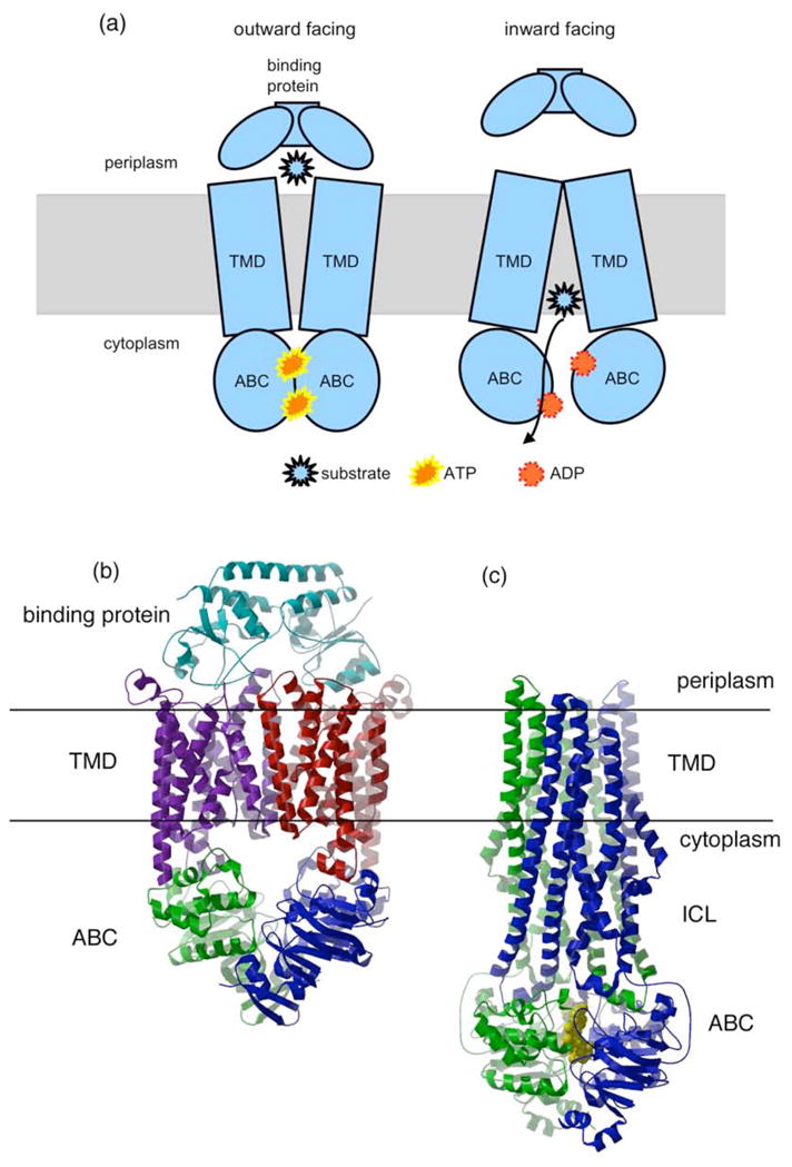
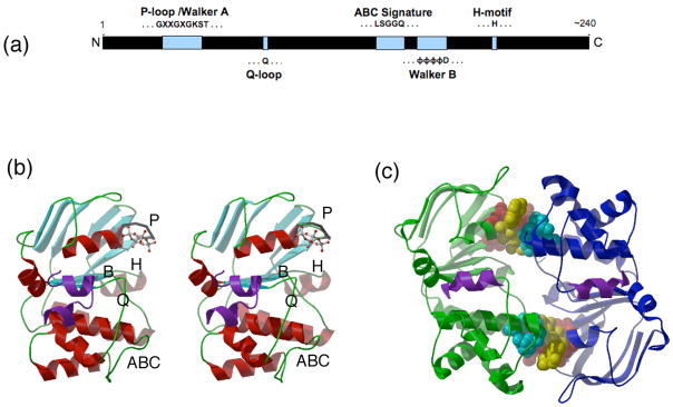
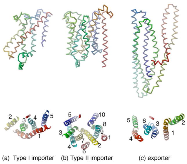
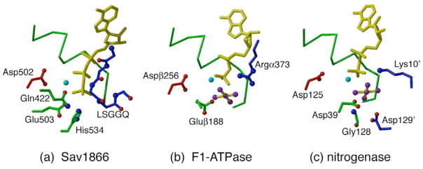
ATP-binding cassette (ABC) transporters constitute a ubiquitous superfamily of integral membrane proteins that are responsible for the ATP-powered translocation of many substrates across membranes. The highly conserved ABC domains of ABC transporters provide the nucleotide-dependent engine that drives transport. By contrast, the transmembrane domains that create the translocation pathway are more variable. Recent structural advances with prokaryotic ABC transporters have provided a qualitative molecular framework for deciphering the transport cycle. An important goal is to develop quantitative models that detail the kinetic and molecular mechanisms by which ABC transporters couple the binding and hydrolysis of ATP to substrate translocation.


Conflict of interest statement

Competing interests statement

The authors declare no competing financial interests

Figures

Figure 1
Figure 1
Molecular architecture of ABC transporters. (a) A cartoon representation of the modular organization of ABC transporters, composed of two transmembrane domains (TMD) and two ABC domains. The binding protein component required by importers is also illustrated. Two conformational states of the ABC transporter, outward facing and inward facing, with the substrate binding site oriented towards the periplasmic (extracellular) and cytoplasmic (intracellular) regions, respectively, are depicted to schematically illustrate the alternating access mechanism of transport (Box 1). (b) The E. coli BtuCDF importer (; PDB 2QI9). The core transporter consists of four subunits, the two membrane spanning subunits, BtuC (purple and red) and the two ABC subunits, BtuD (green and blue). This complex also contains one copy of BtuF, the periplasmic binding protein (cyan). (c) The S. aureus Sav1866 multidrug exporter (; PDB 2ONJ). Sav1866 consists of two subunits (green and blue), which contain a fused TMD and NBD. The bound nucleotides in this structure are represented by yellow space filling models. The periplasmic and cytoplasmic surfaces oriented towards the top and bottom of the figure, respectively. Molecular figures in this article were prepared with MOLSCRIPT and RASTER3D, using coordinates from the Protein Data Bank (PDB77).
Figure 2
Figure 2
Structure and dimer interactions of an ABC subunit. (a) A linear representation of the protein sequence of an ABC domain, illustrating the relative positions along the polypeptide chain of the conserved amino acid motifs. (b) Stereoview of the ABC subunit BtuD (; PDB 1L7V). The P-loop, Walker B, Q-loop, H-motif and ABC signature motif, labeled as ‘P’, ‘B’, ‘Q’, ‘H’ and ‘ABC’, respectively, are positioned along one surface of the subunit. A cyclotetravandate bound to the P-loop is shown as a ball-and-stick model. (c) The nucleotide mediated ABC dimer from Sav1866 (; PDB 2ONJ). The two NBDs are represented as ribbons (green and blue), while the sandwiched AMPPNP nucleotides are in yellow space filling models. The P-loops and the signature motifs are depicted in red and cyan space filling models, respectively. The coupling helices of the TMDs in contact with the ABCs are shown as purple ribbons. The view is down the molecular two-fold axis.
Figure 3
Figure 3
The polypeptide folds of a single transmembrane domain from representative ABC transporters. (a) The Type I ABC importer fold of MetI (PDB 3DHW), (b) the Type II ABC importer fold of BtuC (PDB 1L7V), (c) the ABC exporter fold of Sav1866 (PDB 2ONJ). At the top are the traces of the polypeptide fold for each structure, illustrated with a color gradient ranging from red at the N-terminus through yellow and green to blue at the C-terminus. The molecular two-fold axis (corresponding to the normal to the membrane plane) is vertical. At the bottom are ribbons representations of the TM helices in these structures, viewed from the periplasmic surface rotated 90° from the top view. The position of the molecular two-fold axis is depicted in red in the lower figures. In (a), all the TM helices (TM1-5) are represented for MetI; in (b) the BtuC helices are divided into two groups, TM1-5 and TM6-10; and in (c) the Sav1866 helices are divided into groups TM1-3 and TM4-6. Within each set, the helices are colored red to blue proceeding along the polypeptide chain from the N- to C-terminus to illustrate the internal symmetry present in BtuC and Sav1866. These internal repeats are related by a rotation axis oriented approximately vertically in the plane of the figure.
Figure 4
Figure 4
Nucleotide-protein interactions in the active sites of various ATPases. (a) AMPPNP bound to the ABC transporter Sav1866 (; PDB 2ONJ), (b) the ADP-AlF4 site of the F1-ATPase (, PDB 1H8E), (c) the ADP-AlF4 state of the nitrogenase Fe-protein (, PDB 1M34). The nucleotide is depicted with yellow bonds, while the Mg+2 (or equivalent) is cyan and the AlF4 (when present) has purple atoms. The P-loop is represented by the green Cα trace at the back of each structure, while the Walker B Asp from the same subunit is shown with red bonds on the left. In Sav1866 (a), potential catalytic residues are Glu503 (adjacent to the Walker B), Gln422 in the Q-loop and His534 of the H-motif. The backbone atoms of the LSGGQ signature motif provided from the dimer-related ABC subunit are depicted with blue bonds. For the F1-ATPase (b), the catalytic residue Gluβ188 occupies the same spatial location as the Q-loop, while Argα373 from the adjacent subunit coincides with the signature motif. For nitrogenase (c), Asp39 in the Switch I region, and Gly128 correspond to the Q-loop and H-motif region, respectively; the catalytic residue Asp129 from the adjacent subunit has no obvious counterpart in the ABC transporters. Lys10 from the adjacent subunit is positioned similarly to the signature motif in the ABC transporters.
Figure 5
Figure 5
Relationships between dimeric ABC structures. The polypeptide fold of BtuD (PDB 1L7V) is shown with the two subunits traced in different shades of blue. The positions of the P-loop and ABC signature motifs (defined by the Cα positions of Gly38 and Gly129, respectively) in BtuD are denoted by the smaller and larger blue spheres, respectively. Following the superposition onto the right-most BtuD (subunit 1) of one ABC domain of Sav1866 (red, 2ONJ), MalK (yellow, 1Q12), HI1470 (cyan; 2NQ2); and MetN (green, 3DHW), the positions of the P-loop and ABC signature motifs in these structures are designated by the appropriately colored spheres. The bound AMPPNPs at the ABC dimer interface of Sav1866 (PDB 2ONJ) are shown in purple. The closed interface characteristic of the ATP bound state, as observed in Sav1866 and MalK, is associated with an intersubunit separation between the P-loop and ABC signature motifs of ~11 Å; in the nucleotide-free structures, the separation increases from 14 Å in BtuD to 16 Å in HI1470 to ~28 Å in MetN. The large separation between ABC subunits in MetN likely underlies the phenomenon of transinhibition, in which methionine binding to a regulatory domain of MetI stabilizes an ATPase inactive form by sterically preventing the formation of the catalytically essential closed interface between these domains.

References

    1. Monod J. Recherches sur la croissance des culture bactériennes. Hermann, Editeurs des Sciences et des Arts; Paris: 1942.
    1. Phillips R, Quake SR. The biological frontier of physics. Physics Today. 2006;59:38–43.
    1. Blattner FR, et al. The complete genome sequence of Escherichia coli K-12. Science. 1997;277:1453–1462. - PubMed
    1. Busch W, Saier MHJ. The transporter classification (TC) system. Crit Rev Biochem Mol Biol. 2002;27:287–337. - PubMed
    1. Stouthamer AH. The search for correlation between theoretical and experimental growth yields. Int Rev Biochem. 1979;21:1–47.

Publication types

Substances