. 2009;61(2):251-64.
doi: 10.1080/01635580802398430.

Differential effects of selenium on benign and malignant prostate epithelial cells: stimulation of LNCaP cell growth by noncytotoxic, low selenite concentrations

Affiliations

Differential effects of selenium on benign and malignant prostate epithelial cells: stimulation of LNCaP cell growth by noncytotoxic, low selenite concentrations

Nur Ozten Kandaş et al. Nutr Cancer. 2009.

Abstract

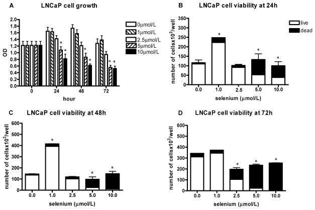
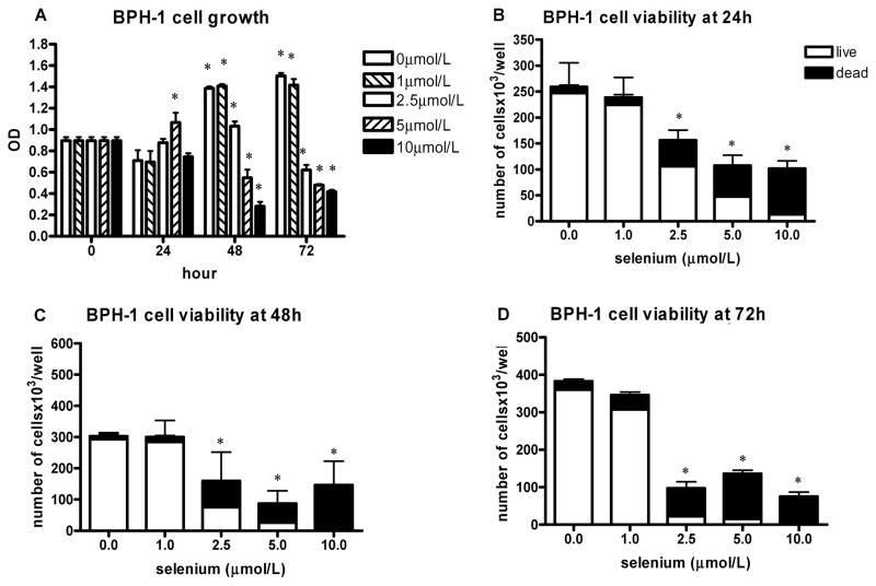
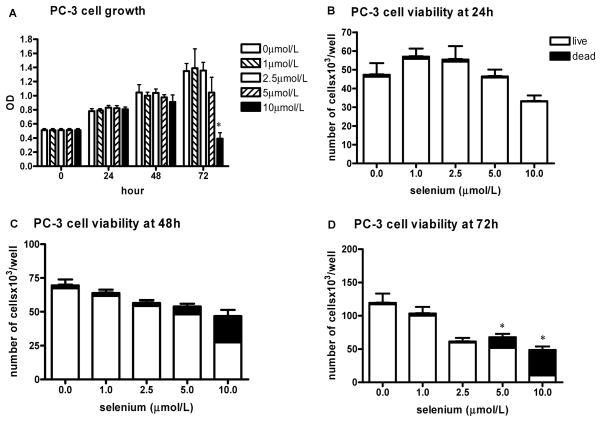
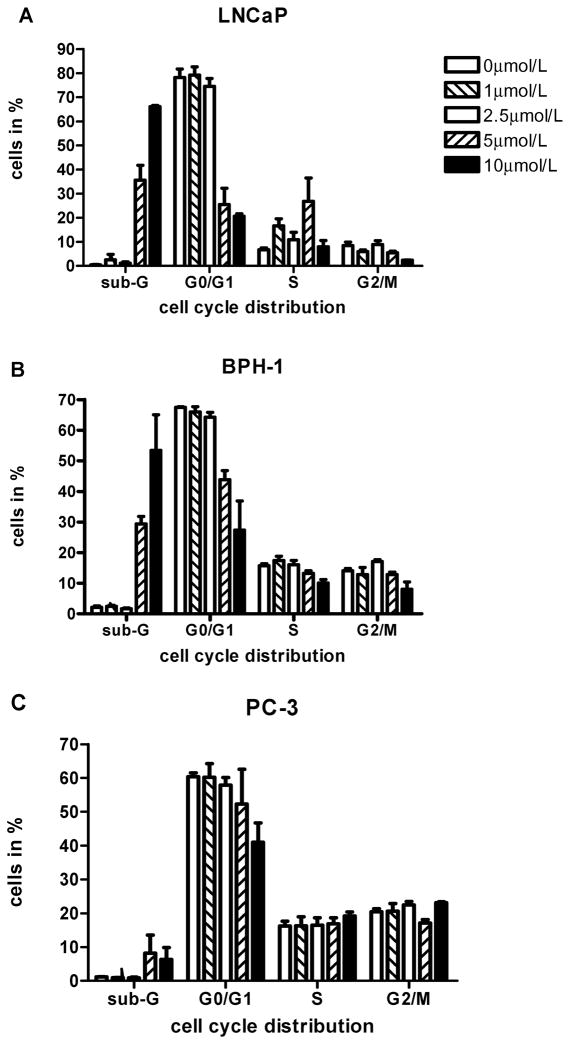
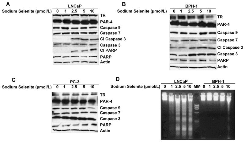
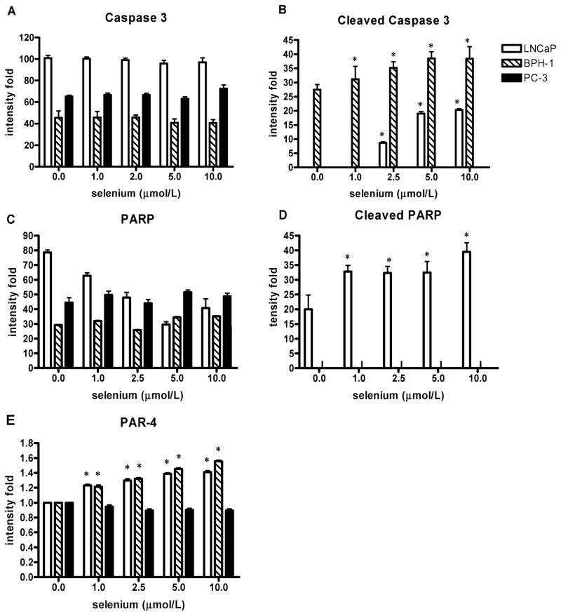
We examined the hypothesis that nontoxic concentrations of selenium induce apoptosis and growth inhibition selectively in prostate cancer cells but not in benign prostate cells. Nontumorigenic BPH-1 prostate epithelial cells, androgen-sensitive LNCaP, and androgen-independent PC-3 prostate cancer cells were exposed to sodium selenite at 1 to 10 micromol/l for 24 to 72 h. Cell proliferation, viability, and apoptosis were assessed by MTT assay, trypan blue exclusion, flow cytometry, DNA laddering, and caspase activation. BPH-1 cells were more sensitive for cytotoxic selenium effects than malignant prostate cells, whereas LNCaP cells were more sensitive than PC-3 cells. At noncytotoxic selenium concentrations, there was no apoptosis in BPH-1 and PC-3 cells and no growth inhibition of LNCaP and BPH-1 cells. PC-3 cells were refractory to apoptosis induction but were growth inhibited at noncytotoxic concentrations. LNCaP cells were growth stimulated at 1 micromol/l and sensitive to apoptosis induction at higher noncytotoxic concentrations. Thus, noncytotoxic selenite concentrations did not induce growth inhibition or apoptosis selectively in prostate cancer cells. Growth stimulation of LNCaP cells by low concentrations suggests the possibility of adverse effects of selenium supplementation on hormone sensitive prostate cancer, whereas inhibition of PC-3 cell proliferation at noncytotoxic concentrations suggests potential benefit of selenium in advanced prostate cancer.


Figures

FIG. 1
FIG. 1
The effect of sodium selenite on LNCaP cell growth and viability. The data represent the mean ± SD of three experiments. A: Cell growth measured by the MTT assay presented as absorbance read by microplate reader at 570 nm at different time intervals; *P < 0.05 (two-sided) for difference with control cells. OD, optical density. B–D: The number of viable cells and cell viability determined by hemocytometer counting and trypan blue exclusion after 24 h (B), 48 h (C), and 72 h (D); *P < 0.001 for difference with control cells. Where error bars are not visible, the variation was very small.
FIG. 2
FIG. 2
The effect of sodium selenite on BPH-1 cell growth and viability. The data represent the mean ± SD of three experiments. A: Cell growth measured by the MTT assay presented as absorbance read by microplate reader at 570 nm at different time intervals; *P < 0.001 (two-sided) for difference with control cells. OD, optical density. B–D: The number of viable cells and cell viability determined by hemocytometer counting and trypan blue exclusion after 24 h (B), 48 h (C), and 72 h (D); *P < 0.001 (two-sided) for difference with control cells.
FIG. 3
FIG. 3
The effect of sodium selenite on PC-3 cell growth and viability. The data represent the mean ± SD of three experiments. A: Cell growth measured by the MTT assay presented as absorbance read by microplate reader at 570 nm at different time intervals; *P < 0.001 (two-sided) for difference with control cells. OD, optical density. B–D: The number of viable cells and cell viability determined by hemocytometer counting and trypan blue exclusion after 24 h (B), 48 h (C), and 72 h (D); *P < 0.001 (two-sided) for difference with control cells.
FIG. 4
FIG. 4
Panels represent summaries of the results of three separate analyses (means ± SD) of cell cycle distribution (sub-G0/G1, G0/G1, S, and G2/M phase, respectively) after treatment of increasing concentrations of sodium selenite (0, 1, 2.5, 5, and 10 μmol/l) for 48 h in A) LNCaP cells, B) BPH-1 cells, and C) PC-3 cells. Where error bars are not visible, the variation was very small.
FIG. 5
FIG. 5
A–C: Western blot analysis of the effect of sodium selenite on cleavage of PARP and selected pro-caspases after 24 h exposure to increasing concentrations of sodium selenite (0, 1, 2.5, 5, and 10 μmol/l) in A) LNCaP cells, B) BPH-1 cells, and C) PC-3 cells. D) Agarose (1.5%) gel electrophoretic detection of nucleosomal DNA fragmentation (laddering) in LNCaP (left 5 lanes) and BPH-1 cells (right 5 lanes) after 72 h exposure to increasing concentrations of sodium selenite. The middle lane, which is indicated as MM, was loaded with 100-bp DNA size markers. TR, thioredoxin reductase; CI, cleaved; PAR-4, Prostate apoptosis response-4.
FIG. 6
FIG. 6
A–E: Effects of sodium selenite treatment of LNCaP, BPH-1, and PC-3 cells for 24 h on caspase 3 and PARP cleavage and Prostate apoptosis response-4 (PAR-4 protein expression. A: Caspase 3. B: Cleaved caspase 3. C: PARP. D: Cleaved PARP. E: PAR-4. Data are expressed as intensity and represent the means ± SD of three independent experiments; *P < 0.05 (two-sided) for difference with control cells. Where error bars are not visible, the variation was very small; and where bars are not visible, there was no detectable protein.

References

    1. Platz EA, Giovannucci E. The epidemiology of sex steroid hormones and their signaling and metabolic pathways in the etiology of prostate cancer. J Steroid Biochem Mol Biol. 2004;92:237–253. [Epub 2005 January 5] - PubMed
    1. Bosland MC. The role of steroid hormones in prostate carcinogenesis. J Natl Cancer Inst Monogr. 2000;27:39–66. - PubMed
    1. Klein EA. Chemoprevention of prostate cancer. Ann Rev Med. 2006;57:49–63. - PubMed
    1. Bosland MC, McCormick DL, Melamed J, Walden PD, Zeleniuch-Jacquotte A, et al. Chemoprevention strategies for prostate cancer. Eur J Cancer Prev. 2002;11(2 Suppl):S18–S27. - PubMed
    1. Sinha R, El-Bayoumy K. Apoptosis is a critical cellular event in cancer chemoprevention and chemotherapy by selenium compounds. Curr Cancer Drug Targets. 2004;4:13–28. - PubMed

Publication types

MeSH terms