Comparative Study
. 2009 Mar;61(3):525-32.
doi: 10.1002/mrm.21808.

Evaluation of the ERETIC method as an improved quantitative reference for 1H HR-MAS spectroscopy of prostate tissue

Affiliations
Comparative Study

Evaluation of the ERETIC method as an improved quantitative reference for 1H HR-MAS spectroscopy of prostate tissue

Mark J Albers et al. Magn Reson Med. 2009 Mar.

Abstract

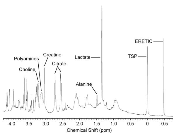
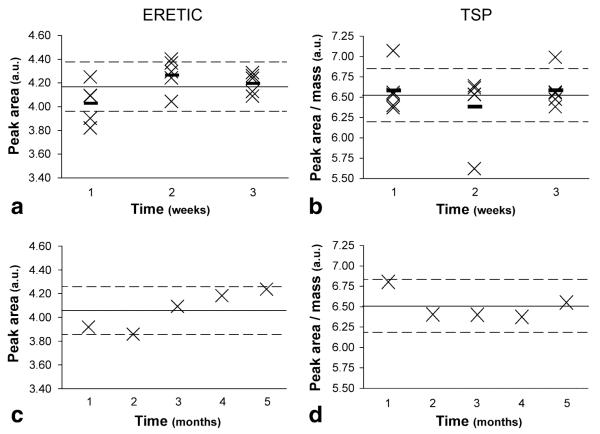
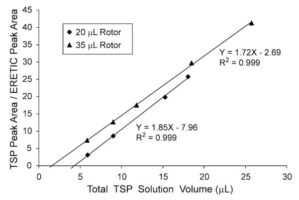
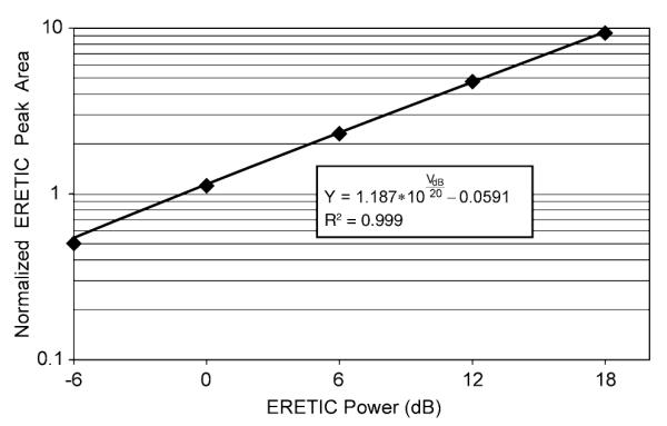
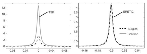
The Electronic REference To access In vivo Concentrations (ERETIC) method was applied to (1)H HR-MAS spectroscopy. The accuracy, precision, and stability of ERETIC as a quantitative reference were evaluated in solution and human prostate tissue samples. For comparison, the reliability of 3-(trimethylsilyl)propionic-2,2,3,3-d(4) acid (TSP) as a quantitation reference was also evaluated. The ERETIC and TSP peak areas were found to be stable in solution over the short-term and long-term, with long-term relative standard deviations (RSDs) of 4.10% and 2.60%, respectively. Quantification of TSP in solution using the ERETIC peak as a reference and a calibrated, rotor-dependent conversion factor yielded results with a precision < or =2.9% and an accuracy error < or =4.2% when compared with the expected values. The ERETIC peak area reproducibility was superior to TSP's reproducibility, corrected for mass, in both prostate surgical and biopsy samples (4.53% vs. 21.2% and 3.34% vs. 31.8%, respectively). Furthermore, the tissue TSP peaks exhibited only 27.5% of the expected area, which would cause an overestimation of metabolite concentrations if used as a reference. The improved quantification accuracy and precision provided by ERETIC may enable the detection of smaller metabolic differences that may exist between individual tissue samples and disease states.


Figures

FIG. 1
FIG. 1
(a) A photograph and (b) a schematic of a wide-mouth zirconia HR-MAS rotor (not drawn to scale). The wide-mouth zirconia rotors were designed to keep the sample within a homogenous region of the RF coil. Although this was true for tissue samples, a small volume, referred to as the invisible volume (VI), of an aqueous sample was forced into the narrow gap between the walls of the rotor bottom and shaft of the plug when the rotor was spinning. Since this volume was outside of the RF coil, it was not detected by the HR-MAS experiment. Using Eq. [3], it was possible to correct for this invisible volume when calibrating the ERETIC signal with a solution sample.
FIG. 2
FIG. 2
(a) Pulse sequence diagram of the ERETIC sequence and (b) system diagram of the Varian INOVA console high-lighting the modifications required for generating an ERETIC signal at the 1H frequency. The output of the 1H synthesizer was shared between the waveform generators (WG) for channels 1 and 3. The ERETIC signal was taken directly from the output of the Programmable Attenuator (PA) module, combined with the output from channel 2 using a directional coupler, and transmitted through the X channel of the nanoprobe. The transmission of the ERETIC waveform and the data acquisition were synchronized in the pulse sequence. (d1, relaxation delay; satdly, presaturation delay; pw, pulse width; at, acquisition time; BP, bandpass).
FIG. 3
FIG. 3
Representative HR-MAS spectrum of prostate surgical tissue with the ERETIC signal generated at a frequency of −0.5 ppm and with a transmitter power of 0 dB. The phase of the ERETIC signal was matched to the metabolites before the acquisition and produced a peak that had a phase and amplitude comparable to those of the tissue metabolites. The frequency of the ERETIC signal was well separated from the other peaks in the spectrum and allowed for accurate quantification of its peak.
FIG. 4
FIG. 4
ERETIC peak area and TSP peak area (corrected for mass) from identically prepared solution samples acquired (a and b) onfive consecutive days over three separate weeks and (c and d) over five months. The solid line represents the mean peak area over the relevant time period, the dotted lines represent 5% deviations from the mean, and the small dashes in plots a and b represent the weekly means. Samples were prepared by adding 3 μL of D2O + TSP (weighed to 0.01 mg) and filling the remainder of the rotor volume with plain D2O. The average weekly RSD of the ERETIC peak area was 4.25%, 3.29%, and 2.09% during each week, respectively, whereas the average relative standard deviation (RSD) of the TSP peak area was 4.37%, 6.76%, and 3.57% during the same periods. The average monthly RSD was 4.10% and 2.60% for ERETIC and TSP, respectively.
FIG. 5
FIG. 5
Plot of the TSP peak area (ATSP)/ERETIC peak area (AE) versus the total volume of the TSP solution determined by weight in the 20 and 35 μL rotors. A linear regression was performed on the data to calculate the rotor-specific conversion factors (ME) and the HR-MAS invisible volumes (VI) contained in Eq. [3]. ME represents the equivalent amount of protons for the ERETIC peak and can be used for quantification of metabolites; VI is the volume of the solution not detected by the HR-MAS experiment. The VI for the 20 μL rotor was 176% greater than the VI for the 35 μL rotor because of small differences in the rotor geometries.
FIG. 6
FIG. 6
A plot of the linearity of ERETIC signal area with ERETIC power. The diamonds represent the average of three HR-MAS measurements taken on the same solution sample after they were normalized to remove arbitrary scaling factors introduced by the spectrometer’s electronics. The black line is a plot of the fit obtained on the data from Matlab. Since the leading coefficient of the fitted model is 1.187, the model indicates that the ERETIC signal area does not exhibit the one-to-one correlation with the ERETIC power that would be expected from an ideal spectrometer.
FIG. 7
FIG. 7
Comparison of the (a) TSP and (b) ERETIC peaks from a prostate surgical sample and solution sample with a comparable amount of TSP. The surgical sample was amplified by 22.1% to correct for loading differences between the two spectra, determined by the differences in the ERETIC peak area and the 90° pulse width. The TSP peak from the solution spectrum was apodized by 2.9 Hz to match the linewidths of the TSP peaks in the two spectra. Since the TSP peak area in the tissue spectrum was 28.7% of the peak area in the solution spectrum, a significant amount of the TSP was not detected in the HR-MAS experiment.

References

    1. Cheng LL, Chang IW, Louis DN, Gonzalez RG. Correlation of high-resolution magic angle spinning proton magnetic resonance spectroscopy with histopathology of intact human brain tumor specimens. Cancer Res. 1998;58:1825–1832. - PubMed
    1. Millis K, Weybright P, Campbell N, Fletcher JA, Fletcher CD, Cory DG, Singer S. Classification of human liposarcoma and lipoma using ex vivo proton NMR spectroscopy. Magn Reson Med. 1999;41:257–267. - PubMed
    1. Swanson MG, Vigneron DB, Tabatabai ZL, Males RG, Schmitt L, Carroll PR, James JK, Hurd RE, Kurhanewicz J. Proton HR-MAS spectroscopy and quantitative pathologic analysis of MRI/3D-MRSI-targeted postsurgical prostate tissues. Magn Reson Med. 2003;50:944–954. - PubMed
    1. Taylor JL, Wu CL, Cory D, Gonzalez RG, Bielecki A, Cheng LL. High-resolution magic angle spinning proton NMR analysis of human prostate tissue with slow spinning rates. Magn Reson Med. 2003;50:627–632. - PubMed
    1. Swanson MG, Zektzer AS, Tabatabai ZL, Simko J, Jarso S, Keshari KR, Schmitt L, Carroll PR, Shinohara K, Vigneron DB, Kurhanewicz J. Quantitative analysis of prostate metabolites using 1H HR-MAS spectroscopy. Magn Reson Med. 2006;55:1257–1264. - PubMed

Publication types