. 2009 Apr;42(2):132-40.
doi: 10.1111/j.1365-2184.2008.00578.x. Epub 2009 Feb 24.

Human immature dental pulp stem cells' contribution to developing mouse embryos: production of human/mouse preterm chimaeras

Affiliations

Human immature dental pulp stem cells' contribution to developing mouse embryos: production of human/mouse preterm chimaeras

S A Siqueira da Fonseca et al. Cell Prolif. 2009 Apr.

Abstract

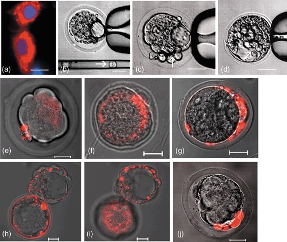
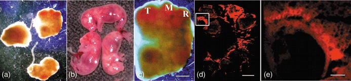
Objectives: In this study, we aimed at determining whether human immature dental pulp stem cells (hIDPSC) would be able to contribute to different cell types in mouse blastocysts without damaging them. Also, we analysed whether these blastocysts would progress further into embryogenesis when implanted to the uterus of foster mice, and develop human/mouse chimaera with retention of hIDPSC derivates and their differentiation.

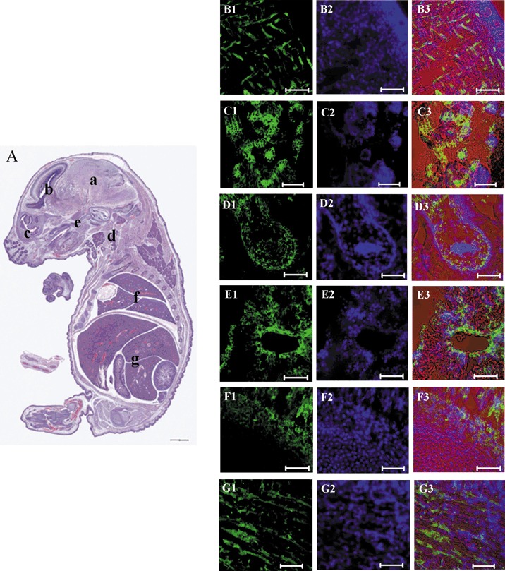
Materials and methods: hIDPSC and mouse blastocysts were used in this study. Fluorescence staining of hIDPSC and injection into mouse blastocysts, was performed. Histology, immunohistochemistry, fluorescence in situ hybridization and confocal microscopy were carried out.

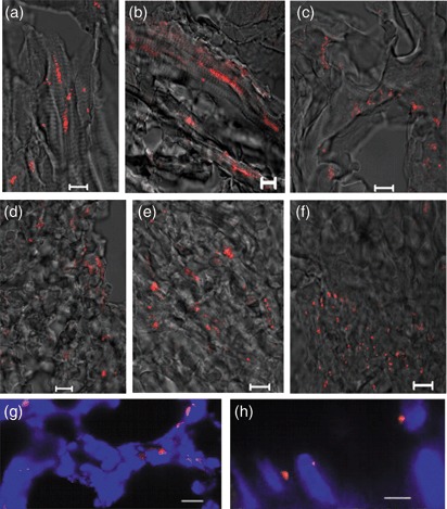
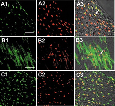
Results and conclusion: hIDPSC showed biological compatibility with the mouse host environment and could survive, proliferate and contribute to the inner cell mass as well as to the trophoblast cell layer after introduction into early mouse embryos (n = 28), which achieved the hatching stage following 24 and 48 h in culture. When transferred to foster mice (n = 5), these blastocysts with hIDPSC (n = 57) yielded embryos (n = 3) and foetuses (n = 6); demonstrating presence of human cells in various organs, such as brain, liver, intestine and hearts, of the human/mouse chimaeras. We verified whether hIDPSC would also be able to differentiate into specific cell types in the mouse environment. Contribution of hIDPSC in at least two types of tissues (muscles and epithelial), was confirmed. We showed that hIDPSC survived, proliferated and differentiated in mouse developing blastocysts and were capable of producing human/mouse chimaeras.


Figures

Figure 1
Figure 1
Human immature dental pulp stem cells (hIDPSC) injection into morulae and blastocysts. (a) hIDPSC stained with Vybrant Cell‐Labelling (red) and nuclei stained with DAPI (blue); (b) hIDPSC before injection (arrow) into blastocysts; (c,d) hIPSC injected into perivitelline space and blastocele, respectively; (e) morula with hIDPSC (red) 24 h after injection; (f) contribution of hIDPSC (red) to the inner cell mass and trophoblast cell layer; (g) hIDPSC restricted to the periviteline space, indicating a lack of migration into blastocele; (h,i) hatching stage of mouse blastocyst with hIDPSC (red), 48 h after injection, showing the presence of these cells in throphoblast in different confocal microscope‐captured sections; (j) mouse fibroblast stained with Vybrant Cell‐Labelling (red) injected into morulae, after 48 h, presenting larger cell morphology than recipient morulae cells. These cells did not integrate into compacted morulae but inhibited progress into blastocyst. Scale bars: a–j = 20 µm. a = epifluorescence; b–d = phase contrast; e–j = merged image of fluorescent confocal microscopy and differential interference contrast.
Figure 2
Figure 2
Mouse embryo with human immature dental pulp stem cells (hIDPSC) at 11 and 18 d.p.c. (a,b) 11 d.p.c. embryos and 18 d.p.c. well‐formed mouse foetuses with hIDPSC, respectively. (c) General aspect of the embryo where the neurosystem primary vesicles is evident: telencephalum (T) mesencephalum (M) and rombencephalum (R). (d) Whole embryo frozen section. (e) Selected area from (d) showing the presence of hIDPSC in ocular region. Scale bars: c, d = 1000 µm; e = 200 µm; a–c = estereoscopic microscope. (d,e) Fluorescence confocal microscopy.
Figure 3
Figure 3
Mouse foetuses with hIDPSC at 18 d.p.c. (A) Histological section of whole embryo, showing apparently normal body organization. At parasagittal or sagittal section where most of the external features of development, which are similar to those seen in newborn mice are evident. The brain (a) shows structures such as lateral ventricles and part of the chorioid plexus. Sub‐arachoid space is also evident (b). Section of parasagittal orbitary cavity is particularly defined (c). Cervical region presents the following structures including mandibular gland, thyroid cartilage and pharynx (d). Palate ossification is a reference to identify nasopharynx and oropharynx (e). Heart is seen only by its lateral wall evident in a trunk region where lung lobes delimitated cranially by the manubrium sterni and costal cartilage (f). Abdominal cavity shows part of the liver lobes, intestine and bladder. Peritoneal cavity is also evident (g). (B1–G3) Confocal microscopy, positive immunostaining with anti‐hIDPSC antibody (Green B1–G1, fluorescent confocal microscopy), nuclei stained with DAPI (Blue B2–G2, epifluorescence) and merged image of fluorescent confocal microscopy, epifluorescence and differential interference contrast (B3–G3). Red is an artificial colour. (B1–B3) brain, (C1–C3) cervical region, (D1–D3) intestine, (E1–E3) liver, (F1–F3) tail, (G1–G3) muscle tissue. Scale bars: A = 1500 µm; B1–G3 = 50 µm.
Figure 4
Figure 4
FISH analysis using human Y chromosome probe (red) in human/mouse chimaeras. (a,b) muscle fibres; (c) ventricular cavity; (d) liver; (e) heart; (f) ocular cavity (merged image of fluorescent confocal microscopy and differential interference contrast); (g) chimaera tissue, showing nuclear localization of Y chromosome signal (red), nuclei stained with DAPI (blue, epifluorescence) and (h) higher magnification of nuclei. As expected, FISH signals localized to the periphery of nuclei. Scale bars: a–h = 10 µm.
Figure 5
Figure 5
In vivo differentiation of hIDPSC into muscle and epithelial cells. (A1) Positive immunostaining with anti‐HumN antibody (green), (A2) nuclei stained with PI (red), A3) merged image of A1 and A2, arrows indicate human nuclei in yellow as a result of superposition of green and red. (B1) Human myosin‐positive muscle fibres (green), B2) anti‐HumN antibody (red), (B3) merged image of B1 and B2, arrows indicate human nuclei. C1) Anti‐human cytokeratin positive immunostaining (green), C2) positive reaction with anti‐HumN antibody (red), (C3) merged image of C1 and C2. Confocal microscopy: A1–G1, B1–G1 = fluorescent confocal microscopy, C1–G1 = fluorescent confocal microscopy + differential interference contrast. Scale bars: A1–C3 = 20 µm.

References

    1. Doss MX, Koehler CI, Gissel C, Hescheler J, Sachinidis A (2004) Embryonic stem cells: a promising tool for cell replacement therapy. J. Cell. Mol. Med. 8, 465–473. - PMC - PubMed
    1. Evans MJ, Kaufman MH (1981) Establishment in culture of pluripotential cells from mouse embryos. Nature 292, 154–156. - PubMed
    1. Martin GR (1981) Isolation of a pluripotent cell line from early mouse embryos cultured in medium conditioned by teratocarcinoma stem cells. Proc. Natl. Acad. Sci. USA 78, 7634–7638. - PMC - PubMed
    1. Thomson JA, Itskovitz‐Eldor J, Shapiro SS, Waknitz MA, Swiergiel JJ, Marshall VS, Jeffrey M (1998) Jones Embryonic stem cell lines derived from human blastocysts. Science 282, 1145–1147. - PubMed
    1. Smith A (2001) Embryonic stem cells In: Marshak DR, Gardner RL, Gottlieb D, eds. Stem Cell Biology, pp. 205–230, Cold Spring Harbor, NY: Cold Spring; Harbor Laboratory Press.

Publication types

MeSH terms