Review
. 2009 Feb 23:4:7.
doi: 10.1186/1750-1172-4-7.

Corneal dystrophies

Affiliations
Review

Corneal dystrophies

Gordon K Klintworth. Orphanet J Rare Dis. .

Abstract

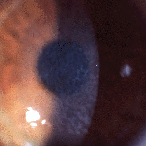
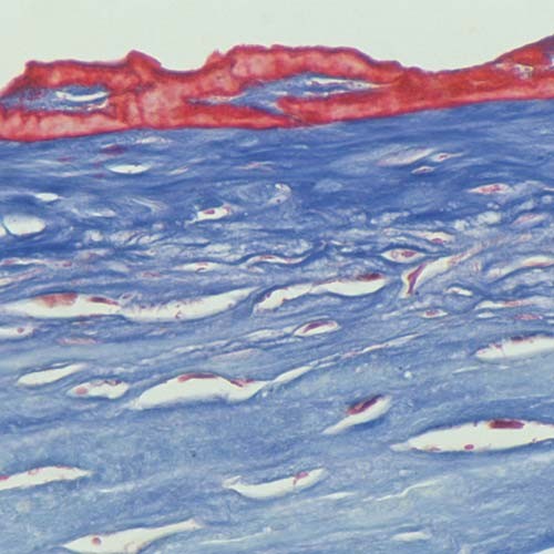
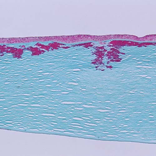
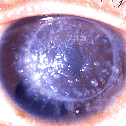
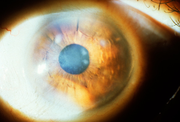
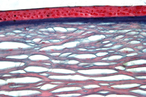
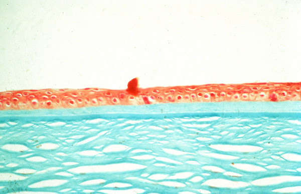
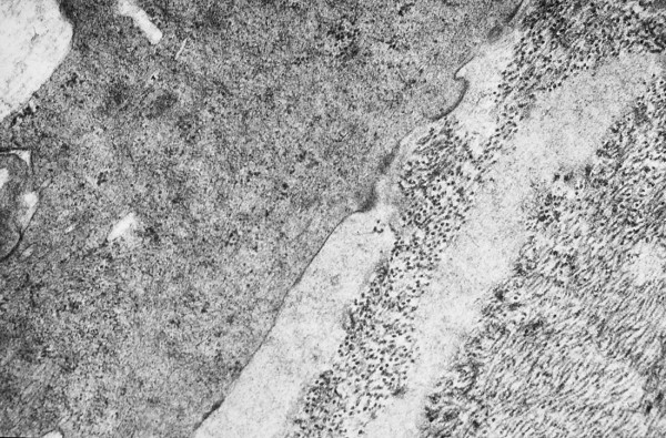
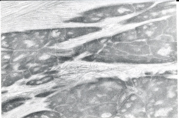
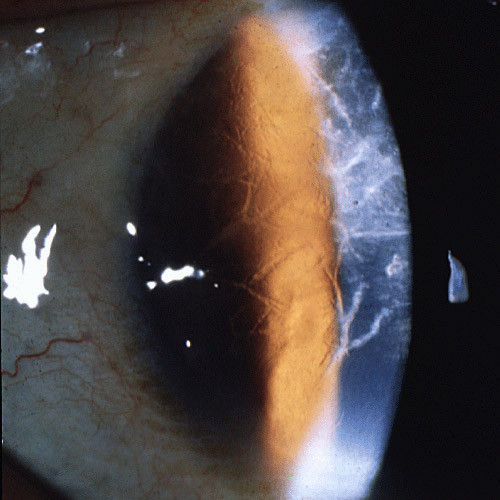
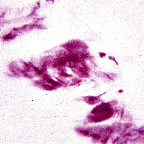
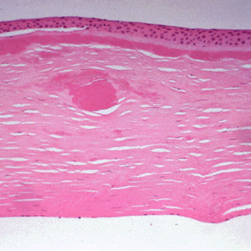
The term corneal dystrophy embraces a heterogenous group of bilateral genetically determined non-inflammatory corneal diseases that are restricted to the cornea. The designation is imprecise but remains in vogue because of its clinical value. Clinically, the corneal dystrophies can be divided into three groups based on the sole or predominant anatomical location of the abnormalities. Some affect primarily the corneal epithelium and its basement membrane or Bowman layer and the superficial corneal stroma (anterior corneal dystrophies), the corneal stroma (stromal corneal dystrophies), or Descemet membrane and the corneal endothelium (posterior corneal dystrophies). Most corneal dystrophies have no systemic manifestations and present with variable shaped corneal opacities in a clear or cloudy cornea and they affect visual acuity to different degrees. Corneal dystrophies may have a simple autosomal dominant, autosomal recessive or X-linked recessive Mendelian mode of inheritance. Different corneal dystrophies are caused by mutations in the CHST6, KRT3, KRT12, PIP5K3, SLC4A11, TACSTD2, TGFBI, and UBIAD1 genes. Knowledge about the responsible genetic mutations responsible for these disorders has led to a better understanding of their basic defect and to molecular tests for their precise diagnosis. Genes for other corneal dystrophies have been mapped to specific chromosomal loci, but have not yet been identified. As clinical manifestations widely vary with the different entities, corneal dystrophies should be suspected when corneal transparency is lost or corneal opacities occur spontaneously, particularly in both corneas, and especially in the presence of a positive family history or in the offspring of consanguineous parents. Main differential diagnoses include various causes of monoclonal gammopathy, lecithin-cholesterol-acyltransferase deficiency, Fabry disease, cystinosis, tyrosine transaminase deficiency, systemic lysosomal storage diseases (mucopolysaccharidoses, lipidoses, mucolipidoses), and several skin diseases (X-linked ichthyosis, keratosis follicularis spinolosa decalvans). The management of the corneal dystrophies varies with the specific disease. Some are treated medically or with methods that excise or ablate the abnormal corneal tissue, such as deep lamellar endothelial keratoplasty (DLEK) and phototherapeutic keratectomy (PTK). Other less debilitating or asymptomatic dystrophies do not warrant treatment. The prognosis varies from minimal effect on the vision to corneal blindness, with marked phenotypic variability.


Figures

Figure 1
Figure 1
Meesmann corneal dystrophy. Multiple opaque spots in the corneal epithelium.
Figure 2
Figure 2
Meesmann corneal dystrophy. Cornea viewed by retroillumination showing numerous small spots.
Figure 3
Figure 3
Meesmann corneal dystrophy. Transmission electron micrograph of the corneal epithelium showing clusters of electrodense fibrillogranular material within a degenerated epithelial cell.
Figure 4
Figure 4
Meesmann corneal dystrophy. Higher magnification transmission electron micrograph of the characteristic peculiar substance (Ps) that is composed of mutated cytokeratin. It is evident in close association with individual filaments (F) (Reproduced with permission from Fine et al.[11]).
Figure 5
Figure 5
Reis-Bücklers corneal dystrophy. Reticular opacity in the superficial cornea.
Figure 6
Figure 6
Reis-Bucklers corneal dystrophy. Light microscopic view of abnormal deposit of fuschinophic mutated transforming growth factor beta induced protein in the superficial corneal strome. Masson trichrome stain (Courtesy of Dr. Guy S. Allaire).
Figure 7
Figure 7
Reis-Bücklers corneal dystrophy. Light microscopy of cornea showing characteristic red stained deposits of mutated transforming growth factor beta induced protein in the superficial corneal stroma. In this specimen they extend deeper into the corneal stroma than the abnormal deposits of Figure 6. Masson trichrome stain.
Figure 8
Figure 8
Gelatinous drop-like corneal dystrophy. A completely opaque cornea with multiple drop-like nodular opacities. Some blood vessels are present in the opaque cornea.
Figure 9
Figure 9
Gelatinous drop-like corneal dystrophy. Recurrent drop-like deposits of subepithelial amyloid are present in the donor tissue of a corneal graft as well as in the surrounding host tissue.
Figure 10
Figure 10
Gelatinous drop-like corneal dystrophy. Light microscopy of subepithelial deposit of amyloid. Hematoxylin and eosin stain.
Figure 11
Figure 11
Gelatinous drop-like corneal dystrophy. Light microscopy view of subepithelial deposit of amyloid. Congo red stain.
Figure 12
Figure 12
Gelatinous drop-like corneal dystrophy. Apple green dichroism of subepithelial deposition of amyloid viewed under polarized light. Congo red stain.
Figure 13
Figure 13
Subepithelial mucinous corneal dystrophy. An irregular shaped opacity is present in the superficial cornea. (Reproduced with permission from Feder et al.[48]).
Figure 14
Figure 14
Subepithelial mucinous corneal dystrophy. A diffuse haziness is present in the papillary region of the cornea in association with discrete opacities. (Reproduced with permission from Feder et al.[48]).
Figure 15
Figure 15
Subepithelial mucinous corneal dystrophy. The region of cornea beneath the epithelium, where Bowman layer is normally present, contains an intensely stained band. Alcian blue stain. (Reproduced with permission from Feder et al.[48]).
Figure 16
Figure 16
Subepithelial mucinous corneal dystrophy. The subepithelial mucoid material stains a lighter green than Bowman layer and the corneal stroma. Masson trichrome stain. (Reproduced with permission from Feder et al.[48])
Figure 17
Figure 17
Subepithelial mucinous corneal dystrophy. Transmission electron microscopic view showing relatively lucent area with fine filaments adjacent to collagen fibers and a keratocyte. (Reproduced with permission from Feder et al.[48])
Figure 18
Figure 18
Macular corneal dystrophy. Discrete opacities are present within hazy cornea that lacks areas of normal clarity.
Figure 19
Figure 19
Macular corneal dystrophy. The abnormalities within the cornea are easily seen within the keratocytes and in a subepithelial extracellular location because they stain prominently with methods that demonstrate glycosaminoglycans. Hale colloidal iron stain.
Figure 20
Figure 20
Macular corneal dystrophy. Transmission electron micrograph of the corneal stroma showing remnant of a keratocyte distended with fibrillogranular material and cellular debris. Some extracellular debris is also present amongst the collagen lamellae (Reproduced with permission from Klintworth [2]).
Figure 21
Figure 21
Macular corneal dystrophy. Transmission electron micrograph of the cytoplasm of a keratocyte showing fibrillogranular material within membrane bound tubules.
Figure 22
Figure 22
Macular corneal dystrophy. Scanning electron micrograph of a section through the corneal stroma showing an accumulation of abnormal material between the collagen lamellae in the location of a keratocyte.
Figure 23
Figure 23
Macular corneal dystrophy. Scanning electron micrograph of the corneal endothelium showing the surface profiles of the nuclei as well as numerous much smaller nodules caused by cytoplasmic accumulations of glycosaminoglycans within the corneal endothelium (Reproduced with permission from Klintworth [1]).
Figure 24
Figure 24
Macular corneal dystrophy. Transmission electron micrograph of the deep corneal stroma, Descemet membrane and the corneal endothelium. This portion of the corneal endothelium contains fibrillogranular material within numerous vacuoles and a distinct vacuolated band is evident in Descemet membrane beneath the most posterior part of this layer.
Figure 25
Figure 25
Macular corneal dystrophy. Higher magnification transmission electron micrograph of the corneal endothelium and Descemet membrane. Note that part of Descemet membrane is associated with small vacuoles and containing osmiophilic material and that the electron density of the portion of Descemet membrane immediately adjacent to the endothelium is variable (Reproduced with permission from Klintworth [1]).
Figure 26
Figure 26
Macular corneal dystrophy. Scanning electron micrograph through a part of Descemet membrane showing a honeycomb appearance due to spaces where abnormal material was lost during tissue processing.
Figure 27
Figure 27
Macular corneal dystrophy. Light microscopic view of a corneal graft in a patient with MCD type I (left side of image) showing a normal brown reactivity of the corneal stroma to the anti-keratan sulfate antibody. The host tissue with MCD type I (right side of image) does not stain. Immunoperoxidase stain with antikeratan sulfate antibody.
Figure 28
Figure 28
Macular corneal dystrophy type I. In contrast to MCD type II (see Figure 29) the corneal deposits do not exhibit antigenic keratan sulfate and hence do not react with antibodies to keratan sulfate. Immunoperoxidase stain with anti-keratan sulfate antibody.
Figure 29
Figure 29
Macular corneal dystrophy type II. The corneal accumulations contain antigenic keratan sulfate and react antibodies to keratan sulfate. Immunoperoxidase stain with antikeratin sulfate antibody.
Figure 30
Figure 30
Granular corneal dystrophy type I. Numerous irregular shaped discrete crumb-like corneal opacities.
Figure 31
Figure 31
Granular corneal dystrophy type II. Variable sized crumb-like opacities in the corneal stroma that have become fused in areas giving rise to elongated and stellate shapes.
Figure 32
Figure 32
Granular corneal dystrophy type II. Photograph of half of a surgically excised piece of cornea showing numerous irregular shaped white corneal opacities that merge with each other.
Figure 33
Figure 33
Granular corneal dystrophy. Light microscopy of cornea showing abnormal eosinophilic deposits in the corneal stroma. Hematoxylin and eosin stain.
Figure 34
Figure 34
Granular corneal dystrophy. Light microscopy of irregular shaped fuchsinophilic (red) deposits in the cornea. Masson trichrome stain.
Figure 35
Figure 35
Granular corneal dystrophy. Higher magnification of characteristic irregular shaped fuchsinophilic (red) deposit in the corneal stroma. Masson trichrome stain.
Figure 36
Figure 36
Granular corneal dystrophy. Image of corneal stroma showing reactivity of the corneal deposits with an antibody to transforming growth factor beta induced protein. Immunoperoxidase stain.
Figure 37
Figure 37
Granular corneal dystrophy. Characteristic rod-shaped bodies in the corneal stroma as seen by transmission electron microscopy. (Reproduced with permission from Klintworth [2]).
Figure 38
Figure 38
Granular corneal dystrophy. Transmission electron micrograph showing moth-eaten appearance of extracellular corneal concretions in deep corneal stroma (Reproduced with permission from Klintworth [2]).
Figure 39
Figure 39
Lattice corneal dystrophy type I. A network of thick linear corneal opacities in patient with a variant of LCD1 (LCD type III) due to a homozygous p. Leu527Arg mutation in the TGFBI gene.
Figure 40
Figure 40
Lattice corneal dystrophy type I. Deposits of amyloid in frozen section of the cornea. Lester King stain.
Figure 41
Figure 41
Lattice corneal dystrophy type I variant. Thicker than usual deposits of eosinophilic amyloid in corneal stroma of patient with a homozygous p. Leu527Arg mutation in the TGFBI gene. Hematoxylin and eosin stain.
Figure 42
Figure 42
Lattice corneal dystrophy type I variant. Deposits of amyloid throughout the corneal stroma due to a p. Ala546Asp mutation in the TFGFBI gene in a patient with a variant of LCD type 1 (polymorphic corneal amyloidosis). (Reproduced with permission from Eifrig et al.[81]).
Figure 43
Figure 43
Lattice corneal dystrophy type I variant. The amyloid within the corneal stroma from a patient with a homozygous p. Leu527Arg mutation in the TGFBI gene viewed under ultraviolet light after staining the fluorescent dye Thioflavin T.
Figure 44
Figure 44
Lattice corneal dystrophy type I. Transmission electron microscopic appearance of amyloid in the upper part of this image adjacent to collagen fibers. (Reproduced with permission from Klintworth [1]).
Figure 45
Figure 45
Lattice corneal dystrophy type II. Diagram depicting gelsolin and the amyloid protein derived from it because of mutations in codon 187 of the GSN gene.
Figure 46
Figure 46
Lattice corneal dystrophy type II. Black and white light micrograph showing deposits of amyloid in cornea. Congo red stain (Reproduced with permission from Klintworth [2]).
Figure 47
Figure 47
Schnyder corneal dystrophy. Crystalline opacities are evident in the central cornea (Courtesy Dr. G.N. Foulks).
Figure 48
Figure 48
Schnyder corneal dystrophy. The central cornea contains crystalline deposits and a prominent opaque ring (annulus lipoides) is evident in the peripheral cornea (Courtesy Dr. Seymour Brownstein).
Figure 49
Figure 49
Schnyder corneal dystrophy. Birefringent crystals of cholesterol are evident in the superficial corneal stroma of this unfixed portion of frozen cornea examined under polarized light. (Courtesy of Dr. M. M. Rodrigues).
Figure 50
Figure 50
Schnyder corneal dystrophy. Transmission electron micrograph showing numerous electron lucent spaces in the cornea caused by dissolved cholesterol crystals (Courtesy of Dr. M. M. Rodrigues) (Reproduced with permission from Klintworth [2]).
Figure 51
Figure 51
Fleck corneal dystrophy. Appearance of the cornea by slit-lamp biomicroscopy (left image) and by confocal microscopy (right image) (Courtesy Dr. Charles N. McGhee).
Figure 52
Figure 52
Fleck corneal dystrophy. The cornea contains small fleck-like opacities and these can be seen by confocal microscopy in the inserts. Note the enlarged cell in the lower insert (Courtesy Dr. Charles N. McGhee).
Figure 53
Figure 53
Congenital stromal dystrophy. The cornea is particularly opaque in the anterior stroma by slit-lamp biomicroscopy (Reproduced with permission from Bredrup et al. [101]).
Figure 54
Figure 54
Congenital stromal dystrophy. Transmission electron microscopy of the corneal stroma showing normal collagen lamellae separated by abnormal randomly distributed collagen filaments in an electron-lucent extracellular matrix (Reproduced with permission from Bredrup et al.[101]).
Figure 55
Figure 55
Fuchs corneal dystrophy. A markedly opaque cornea caused by extensive edema due to a loss of endothelial cells that normal maintain the hydrophilic corneal stroma in a deturgescent state.
Figure 56
Figure 56
Fuchs corneal dystrophy. Light microscopic appearance of the corneal endothelium, Descemet membrane, and the adjacent corneal stroma showing guttae, a paucity of endothelial cells including some containing melanosomes. Hematoxylin and eosin stain.
Figure 57
Figure 57
Fuchs corneal dystrophy. Transmission electron micrograph showing attenuated corneal endothelial cell with markedly electron dense cytoplasm and haphazardly arranged filaments within Descemet membrane.
Figure 58
Figure 58
Fuchs corneal dystrophy. Transmission electron micrograph of a melanosome containing corneal endothelial cell closely adherent to a collagenous layer with fibrils orientated in different directions (Reproduced with permission from Klintworth [2]).
Figure 59
Figure 59
Fuchs corneal dystrophy. View of guttae on inner surface of the cornea by scanning electron microscopy.
Figure 60
Figure 60
Fuchs corneal dystrophy. Transmission electron microscopic view of broad-banded collagen within the thickened Descemet membrane.
Figure 61
Figure 61
Fuchs corneal dystrophy. A subepithelial bulla resulting from a separation of the corneal epithelium from Bowman layer. This bullous keratopathy is a manifestation of numerous disorders of the cornea in which fluid accumulates beneath the corneal epithelium following a functionally defective endothelial layer.
Figure 62
Figure 62
Fuchs corneal dystrophy. Light microscopic appearance of the cornea showing numerous excrescences on the posterior surface of Descemet membrane (guttae) and the presence of cysts in the corneal epithelium beneath ectopically placed intraepithelial basement membrane. Periodic acid-Schiff stain.
Figure 63
Figure 63
Fuchs corneal dystrophy. Higher magnification of the epithelium in Figure 62 illustrating the intraepithelial cysts and ectopic epithelial basement membrane. This non-specific tissue reaction occurs in many disorders affecting the corneal epithelium and is commonly referred to as epithelial basement membrane dystrophy. Periodic acid-Schiff stain.
Figure 64
Figure 64
Posterior polymorphous corneal dystrophy. Appearance of the abnormal corneal endothelial cells that have become transformed into stratified squamous epithelium. Periodic acid Schiff stain. (Courtesy Dr. Ralph C. Eagle, Jr).
Figure 65
Figure 65
Posterior polymorphous corneal dystrophy. Immunohistochemical demonstration of cytokeratin (stained brown) within the transformed corneal endothelium which has acquired the attributes of stratified squamous epithelium. Immunoperoxidase stain with antibody to cytokeratin.
Figure 66
Figure 66
Posterior polymorphous corneal dystrophy. Scanning electron microscopic view of the corneal endothelium showing the surface profiles of endothelial (ENDO) and epithelial cells (EPI) (Reproduced with permission from Rodriques et al. [130]).
Figure 67
Figure 67
Posterior polymorphous corneal dystrophy. Transmission electron micrograph showing epithelial-like cells lining the inner surface of the cornea. (Reproduced with permission from Boruchoff and Kuwabara [131]).
Figure 68
Figure 68
Posterior polymorphous corneal dystrophy. Scanning electron micrograph showing numerous microvilli on the surface of a corneal endothelial cell (Reproduced with permission from Klintworth [2]).
Figure 69
Figure 69
Congenital hereditary endothelial dystrophy. A markedly opaque cornea due to stromal edema secondary to defective endothelial cells (Courtesy of Dr. Ahmed A. Hidajat).
Figure 70
Figure 70
Congenital hereditary endothelial dystrophy type 2. Macroscopic appearance of thick edematous cornea from a patient with CHED2 (left image) compared to control (right specimen) (Courtesy of Dr. Susan Kennedy).
Figure 71
Figure 71
Congenital hereditary endothelial dystrophy. Transmission electron micrograph of Descemet membrane and the corneal endothelium of a patient from an inbred family with CHED2 illustrating haphazardly arranged broad-banded collagen fibers within Descemet membrane (Courtesy of Dr. Susan Kennedy).

References

    1. Klintworth GK. Genetic disorders of the cornea. In: Klintworth GK, Garner A, editor. Garner and Klintworth's Pathobiology of Ocular Disease. Third. New York: Informa Heathcare; 2008. pp. 655–712.
    1. Klintworth GK. Corneal dystrophies. In: Nicholson DH, editor. Ocular Pathology Update. New York: Masson; 1980. pp. 23–54.
    1. Klintworth GK. The molecular genetics of the corneal dystrophies – current status. Frontiers in Bioscience. 2002;8:687–713. doi: 10.2741/1018. - DOI - PubMed
    1. Weiss JS, Moller H, Lisch W, Kinoshita S, Aldave A, Belin MW, Busin M, Kim EK, Munier F, Seitz W, Sutphin J, Bredrup C, Mannis M, Rapuano C, van Rij G, Kim EK, Klintworth GK. The IC3D Classification of Corneal Dystrophies. Cornea. 2008;27:S1–S42. - PMC - PubMed
    1. Szaflik JP, Oldak M, Maksym RB, Kaminska A, Pollak A, Udziela M, Ploski R, Szaflik J. Genetics of Meesmann corneal dystrophy: a novel mutation in the keratin 3 gene in an asymptomatic family suggests genotype-phenotype correlation. Mol Vis. 2008;14:1713–1718. - PMC - PubMed

Publication types

MeSH terms