. 2009 Feb 24;28(1):25.
doi: 10.1186/1756-9966-28-25.

Endoplasmic reticulum Ca2+-homeostasis is altered in Small and non-small Cell Lung Cancer cell lines

Affiliations

Endoplasmic reticulum Ca2+-homeostasis is altered in Small and non-small Cell Lung Cancer cell lines

Albrecht Bergner et al. J Exp Clin Cancer Res. .

Abstract

Background: Knowledge of differences in the cellular physiology of malignant and non-malignant cells is a prerequisite for the development of cancer treatments that effectively kill cancer without damaging normal cells. Calcium is a ubiquitous signal molecule that is involved in the control of proliferation and apoptosis. We aimed to investigate if the endoplasmic reticulum (ER) Ca2+-homeostasis is different in lung cancer and normal human bronchial epithelial (NHBE) cells.

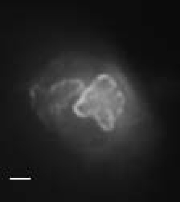
Methods: The intracellular Ca2+-signaling was investigated using fluorescence microscopy and the expression of Ca2+-regulating proteins was assessed using Western Blot analysis.

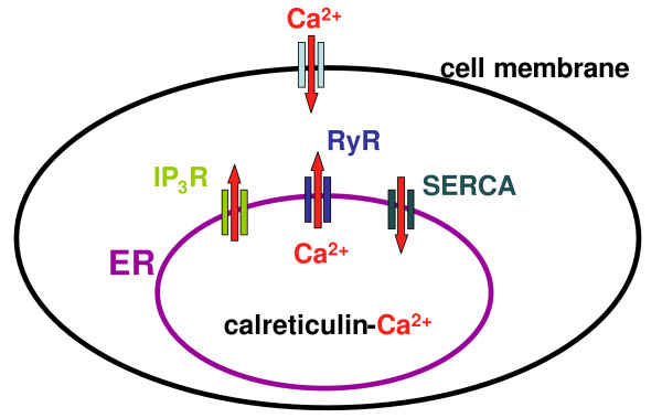
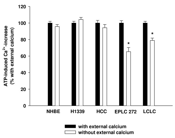
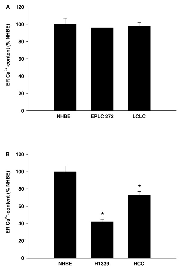
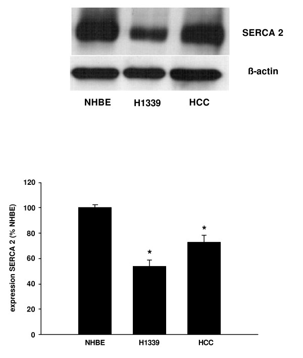
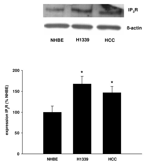
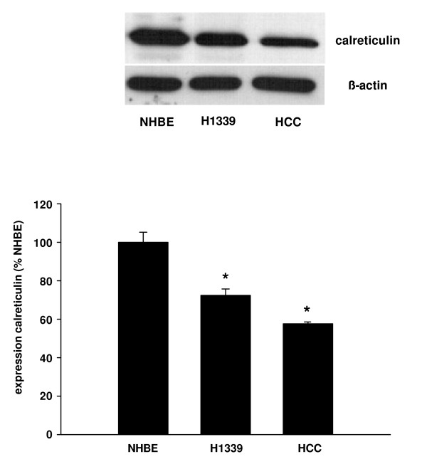
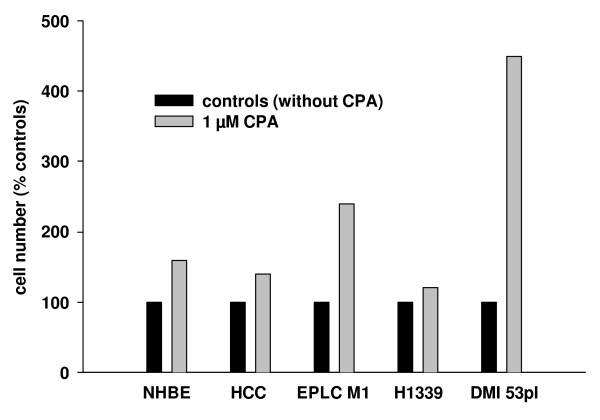
Results: In a Small Cell Lung Cancer (H1339) and an Adeno Carcinoma Lung Cancer (HCC) cell line but not in a Squamous Cell Lung Cancer (EPLC) and a Large Cell Lung Cancer (LCLC) cell line the ER Ca2+-content was reduced compared to NHBE. The reduced Ca2+-content correlated with a reduced expression of SERCA 2 pumping calcium into the ER, an increased expression of IP3R releasing calcium from the ER, and a reduced expression of calreticulin buffering calcium within the ER. Lowering the ER Ca2+-content with CPA led to increased proliferation NHBE and lung cancer cells.

Conclusion: The significant differences in Ca2+-homeostasis between lung cancer and NHBE cells could represent a new target for cancer treatments.


Figures

Figure 1
Figure 1
Increase in the cytoplasmic Ca2+-concentration can be due to Ca2+-influx from the extracellular space or due to Ca2+-release from the endoplasmic reticulum (ER). The equilibrium of the ER Ca2+-content is maintained by sarcoplasmic/endoplasmic reticulum Ca2+-ATPases (SERCA) pumping calcium into the ER and inositol-1,4,5-phosphate- (IP3R) and ryanodine-receptors (RYR) releasing calcium out of the ER. Within the ER, calcium is mainly buffered by calreticulin.
Figure 2
Figure 2
Cells were exposed to 1 mM ATP in the presence and the absence of extracellular calcium. The resulting increase in the cytoplasmic Ca2+-concentration was quantified using fluorescence microscopy. For each cell line, the Ca2+-increase with external calcium was set to 100% (black columns) and the Ca2+-increase without external calcium (white columns) was expressed as percent of the increase with external calcium. In normal bronchial epithelial (NHBE), Small Cell Lung Cancer (H1339) and Adeno-Carcinoma (HCC) cells, the ATP-induced Ca2+-increase was independent of the presence of extracellular calcium suggesting a minor role for Ca2+-influx. In Squamous Cell Lung Carcinoma (EPLC) and Large Cell Lung Carcinoma (LCLC) cells, the ATP-induced Ca2+-increase was lower in the absence of extracellular calcium (n = 42 – 162 cells, * = P < 0.001 versus "with external calcium").
Figure 3
Figure 3
SERCA were inhibited using 1 μM cyclopiazonic acid leading to a net Ca2+-efflux out of the ER. The resulting increase in [Ca2+]c was used as an estimate of the [Ca2+]ER and expressed as percentage of NHBE. (A) In EPLC and LCLC cells in which Ca2+-influx contributed to the ATP-induced Ca2+-increase the [Ca2+]ER was equal to the [Ca2+]ER of NHBE. (B) In H1339 and HCC cells in which the Ca2+-influx did not contribute to the ATP-induced Ca2+-increase the ER Ca2+-content was lower than in NHBE cells (n = 50 – 153 cells, * = P < 0.001 versus all other groups).
Figure 4
Figure 4
Immunohistochemical staining of SERCA 2 in a H1339 cell. Note the ER-typical pattern of the staining as SERCA is an ER-trans-membrane protein. Bar = 2 μm.
Figure 5
Figure 5
The expression of SERCA 2 was analyzed in NHBE, H1339, and HCC cells using Western Blot analysis and expressed as percentage of the SERCA 2 expression in NHBE cells. In H1339 and HCC cells, the expression of SERCA 2 was found to be reduced with H1339 showing the weakest expression (n = 3, * = P < 0.01 versus all other groups).
Figure 6
Figure 6
The expression of IP3R was analyzed in NHBE, H1339, and HCC cells using Western Blot analysis and expressed as percentage of the IP3R expression in NHBE cells. In H1339 and HCC cells, the expression of IP3R was increased with H1339 showing the highest expression (n = 4, * = P < 0.01 versus all other groups).
Figure 7
Figure 7
The expression of calreticulin was analyzed in NHBE, H1339, and HCC cells using Western Blot analysis and expressed as percentage of the calreticulin expression in NHBE cells. In H1339 and HCC cells, the expression of calreticulin was reduced with HCC cells showing the weakest expression (n = 3, * = P < 0.01 versus all other groups).
Figure 8
Figure 8
Cells were treated with 1 μM CPA for 24 h to inhibit SERCA. The cell number was assessed after 24 h and expressed as percent of the non-treated controls. In NHBE cells, non-small cell lung cancer cells (HCC and EPLC M1), and small cell lung cancer cells (H1339 and DMI 53 pI) the cell number was higher after CPA treatment.

References

    1. Alberg AJ, Ford JG, Samet JM. Epidemiology of lung cancer: ACCP evidence-based clinical practice guidelines (2nd edition) Chest. 2007;132:29S–55S. doi: 10.1378/chest.07-1347. - DOI - PubMed
    1. Berridge MJ, Bootman MD, Roderick HL. Calcium signalling: dynamics, homeostasis and remodelling. Nat Rev Mol Cell Biol. 2003;4:517–29. doi: 10.1038/nrm1155. - DOI - PubMed
    1. Clapham DE. Calcium signaling. Cell. 2007;131:1047–58. doi: 10.1016/j.cell.2007.11.028. - DOI - PubMed
    1. Bergner A, Kellner J, Silva AK, Gamarra F, Huber RM. Ca2+-signaling in airway smooth muscle cells is altered in T-bet knock-out mice. Respir Res. 2006;7:33. doi: 10.1186/1465-9921-7-33. - DOI - PMC - PubMed
    1. Wuytack F, Raeymaekers L, Missiaen L. Molecular physiology of the SERCA and SPCA pumps. Cell Calcium. 2002;32:279–305. doi: 10.1016/S0143416002001847. - DOI - PubMed

Publication types

MeSH terms

Substances