Review
. 2009 Aug 1;238(3):240-9.
doi: 10.1016/j.taap.2009.01.028. Epub 2009 Feb 21.

Cadmium-induced testicular injury

Affiliations
Review

Cadmium-induced testicular injury

Erica R Siu et al. Toxicol Appl Pharmacol. .

Abstract

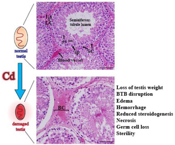
Cadmium (Cd) is an environmental toxicant and an endocrine disruptor in humans and rodents. Several organs (e.g., kidney, liver) are affected by Cd and recent studies have illustrated that the testis is exceedingly sensitive to Cd toxicity. More important, Cd and other toxicants, such as heavy metals (e.g., lead, mercury) and estrogenic-based compounds (e.g., bisphenols) may account for the recent declining fertility in men among developed countries by reducing sperm count and testis function. In this review, we critically discuss recent data in the field that have demonstrated the Cd-induced toxicity to the testis is probably the result of interactions of a complex network of causes. This is likely to involve the disruption of the blood-testis barrier (BTB) via specific signal transduction pathways and signaling molecules, such as p38 mitogen-activated protein kinase (MAPK). We also summarize current studies on factors that confer and/or regulate the testis sensitivity to Cd, such as Cd transporters and metallothioneins, the impact of Cd on the testis as an endocrine disruptor and oxidative stress inducer, and how it may disrupt the Zn(2+) and/or Ca(2+) mediated cellular events. While much work is needed before a unified mechanistic pathway of Cd-induced testicular toxicity emerges, recent studies have helped to identify some of the likely mechanisms and/or events that take place during Cd-induced testis injury. Furthermore, some of the recent studies have shed lights on potential therapeutic or preventive approaches that can be developed in future studies by blocking or minimizing the destructive effects of Cd to testicular function in men.


Figures

Figure 1
Figure 1. Cd-induced testicular injury in the testis
A schematic drawing illustrating a normal testis and a damaged testis after Cd exposure, representing the most common gross morphological changes induced by Cd: hemorrhage and testis weight loss. The histological injury induced by Cd is noticeable when comparing micrographs of hematoxylin-eosin stained paraffin cross-sections of testes from rats terminated after 20 hr of treatment with vehicle (0.9% NaCl; upper panel) or CdCl2 (3 mg/kg b.w., i.p.; lower panel). In normal testis (upper panel), the micrograph illustrates the well-organized distribution of cells in the seminiferous epithelium (SC, Sertoli cell; es, elongating spermatid; sp, spermatocyte); the interstitial Leydig cells (LC) and intact blood vessels in the interstitium. After Cd treatment, disruption of the tight junctions in the microvessels led to the leakage of blood cells (BC, lower panel), most notably erythrocytes, into the interstitial space, causing hemorrhage and edema. In addition, Cd affected Leydig cells (LC), thereby promoting a reduction in steroidogenesis, and Cd also induced cell necrosis, for example, by production of reactive oxygen species. Taken together, these effects induced by Cd exposure result in germ cell loss, causing irreversible sterility. Scale bar, 80 μm.
Figure 2
Figure 2. A schematic drawing illustrating the relative location of the blood-testis barrier (BTB) and various junction types found in the seminiferous epithelium at the Sertoli-Sertoli and/or Sertoli-germ cell interface which are the targets of Cd toxicity in adult rat testes
The seminiferous epithelium, consisted of Sertoli cells and germ cells at different stages of their development, is overlying the tunica propria, which is composed of the non-cellular zone (basement membrane, type I collagen layer) and the cellular zone (peritubular myoid cell layer and the lymphatic vessel). The BTB, created by adjacent Sertoli cells near the basement membrane, divides the seminiferous epithelium into the basal and the adluminal compartments. The BTB is constituted by coexisting tight junction (TJ), desmosome-like junction, gap junction, and basal ectoplasmic specialization (basal ES, which is a testis-specific actin-based atypical adherens junction type), which is one of the primary targets of Cd toxicity in the testis. It is noted that pre/leptotene spermatocytes traverse the BTB at stage VIII-IX of the seminiferous epithelial cycle of spermatogenesis, so that post-meiotic germ cell development takes place behind the BTB, wherein secondary spermatocytes develop into round, elongating and elongated spermatids.
Figure 3
Figure 3. Cellular localization of focal adhesion kinase (FAK) and occludin and their redistribution following CdCl2 treatment in Sertoli cells cultured in vitro
Primary rat Sertoli cells (0.05 × 10⁶ cells/cm2) were cultured on Matrigel-coated coverslips for 4 days, known to form functional tight junction, basal ES and desmosome-like junction, that mimics the BTB in vivo (Siu et al., 2005), and stained for FAK by incubation with mouse monoclonal anti-FAK antibody (Upstate, Cat No.: 05-537, Lot: 0701049384) and goat anti-mouse IgG AlexaFluor 488-conjugated secondary antibody. FAK was localized to the Sertoli-Sertoli cells interface and some staining was found in the cytosol and nuclei (A, a). Treatment with 3 μM CdCl2 caused a redistribution of FAK from the cell-cell interface towards the cytoplasm that was visible after 3 hr (A, b) and more evident after 6 hr (A, c), illustrating a time-dependent effect. The specificity of the FAK antibody used in this study was confirmed by immunoblot using Sertoli cell lysates (30 μg of protein), which shows a single band of ~125 kDa (B). FAK was shown to co-localize with occludin (C, a-c), which is an integral membrane tight junction protein at the BTB, illustrating it is a component of the BTB consistent with an earlier report (Siu et al., 2003). Following CdCl2 treatment (3 μM, 6 hr) both proteins were mis-localized (C, d-f), moving away from the Sertoli-Sertoli cell interface, suggesting that FAK may play a role in the damage of Sertoli-Sertoli cell junctions induced by Cd. Scale bars = 20 μM.
Figure 4
Figure 4. Cd-induced blood-testis barrier (BTB) and cell junction disruption in the seminiferous epithelium is mediated by the activation of specific signal transduction pathways
A schematic drawing illustrating the likely sequence of events in the seminiferous epithelium following Cd treatment (e.g., 3 mg/kg b.w. via i.p.) that leads to BTB disruption and germ cell loss in three phases. In Phase I, Cd ions likely enter Sertoli and/or germ cell via different mechanisms, such as diffusion, Ca+2 channels or using a Cd+2/Zn+2 transporter (Step 1). Although the Cd transporter, ZIP8, is present in Sertoli cells in the mouse, it is not known if it is present in all rodents and/or mammals, as well as in germ cells. Once inside the Sertoli and/or germ cell, Cd induces the synthesis and release of cytokines (e.g., TGFβ-3), which, by interacting with their respective receptors, activates the stress activated p38 MAPK signaling pathway (Step 2) that can selectively disrupt the BTB. At the same time, possibly also mediated by the p38 MAPK, the production of proteases (e.g., cathepsin L) is also induced (Step 3). In Phase II, the BTB is disrupted via degradation of the integral membrane proteins (via the effects of proteases) and/or an acceleration of clathrin-mediated endocytosis of tight junction (e.g., occludin) and basal ES proteins (e.g., N-cadherin). The net results of all this contribute to the disruption of the BTB and Sertoli-germ cell junctions that lead to germ cell loss (Step 4). In Phase III, in order to limit unwanted proteolysis and avoid the dissolution of the entire seminiferous epithelium in the testis, there is an activation of c-JNK signaling pathway, which, in turn, stimulates the production of protease inhibitors (e.g., α2-macroglobulin) to initiate recovery of the seminiferous epithelium (Step 5). This cascade of events depicted herein is based on recent findings in the field as detailed in the text.

References

    1. Acharya UR, Mishra M, Patro J, Panda MK. Effect of vitamins C and E on spermatogenesis in mice exposed to cadmium. Reprod. Toxicol. 2008;25:84–88. - PubMed
    1. Amara S, Abdelmelek H, Garrel C, Guiraud P, Douki T, Ravanat JL, Favier A, Sakly M, Ben Rhouma K. Preventive effect of zinc against cadmium-induced oxidative stress in the rat testis. J. Reprod. Dev. 2008;54:129–134. - PubMed
    1. ATSDR . Agency for Toxic Substances and Disease Registry. U.S. Department of Health and Human Services; Atlanta, GA: 2007. The 2007 CERCLA priority list of hazardous substances.
    1. ATSDR . Agency for Toxic Substances and Disease Registry. U.S. Department of Health and Human Services; Atlanta, GA: 2008. Cadmium toxicity - Case Studies in Environmental Medicine.
    1. Beardsley A, Robertson DM, O'Donnell L. A complex containing a6b1-integrin and phosphorylated focal adhesion kinase between Sertoli cells and elongated spermatids during spermatid release from the seminiferous epithelium. J. Endocrinol. 2006;190:759–770. - PubMed

Publication types

MeSH terms