. 2009 May 5;160(2):264-74.
doi: 10.1016/j.neuroscience.2009.02.025. Epub 2009 Feb 21.

Sensitizing regimens of (+/-)3, 4-methylenedioxymethamphetamine (ecstasy) elicit enduring and differential structural alterations in the brain motive circuit of the rat

Affiliations

Sensitizing regimens of (+/-)3, 4-methylenedioxymethamphetamine (ecstasy) elicit enduring and differential structural alterations in the brain motive circuit of the rat

K T Ball et al. Neuroscience. .

Abstract

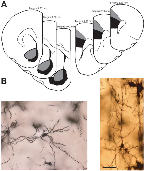
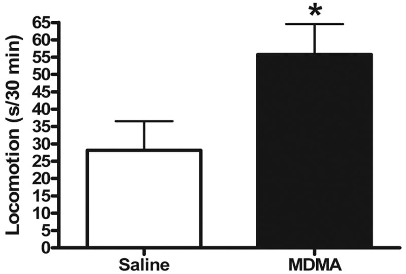
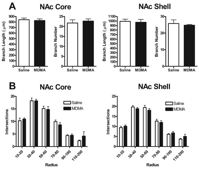
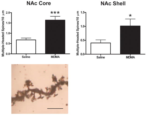
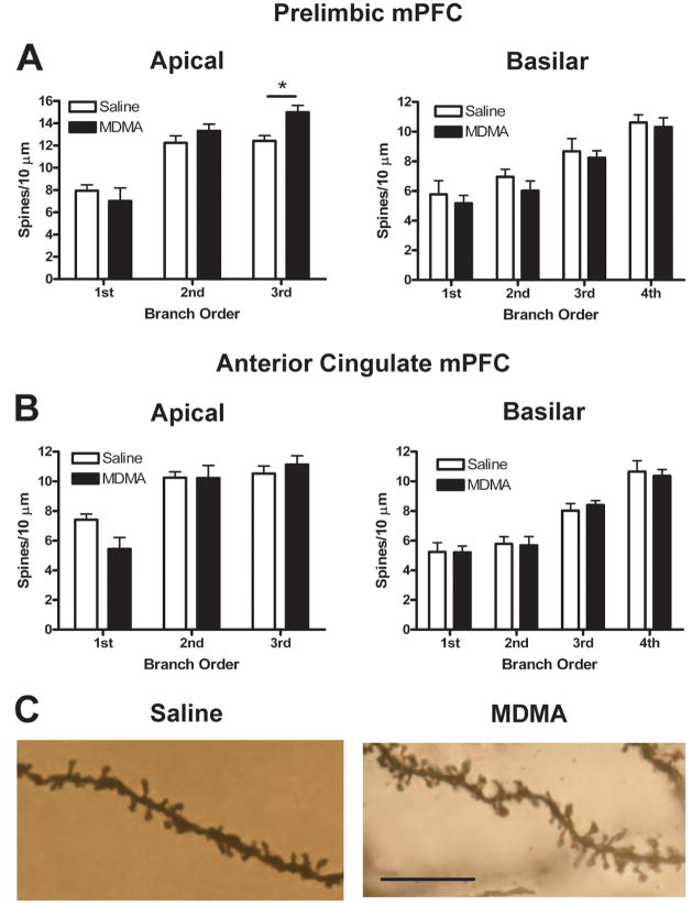
Repeated, intermittent exposure to the psychomotor stimulants amphetamine and cocaine induces a progressive and enduring augmentation of their locomotor-activating effects, known as behavioral sensitization, which is accompanied by similarly stable adaptations in the dendritic structure of cortico-striatal neurons. We examined whether repeated exposure to the increasingly abused amphetamine derivative 3,4-methylenedioxymethamphetamine (MDMA; ecstasy) also results in long-lasting behavioral and morphological changes in mesocortical (medial prefrontal cortex) and ventral striatal (nucleus accumbens) neurons. Rats received two daily injections of either 5.0 mg/kg (+/-)-MDMA or saline vehicle, approximately 6 h apart, for 3 consecutive days, followed by 4 drug-free days for a total of 3 weeks. Following a 4-week drug-free period, MDMA-pretreated rats displayed behavioral sensitization, as well as large increases in spine density and the number of multiple-headed spines on medium spiny neurons in core and shell subregions of nucleus accumbens. In medial prefrontal cortex, the prelimbic subregion showed increased spine density on distal dendrites of layer V pyramidal neurons, while the anterior cingulate subregion showed a change in the distribution of dendritic material instead. Collectively, our results show that long-lasting locomotor sensitization to MDMA is accompanied by reorganization of synaptic connectivity in limbic-cortico-striatal circuitry. The differential plasticity in cortical subregions, moreover, suggests that drug-induced structural changes are not homogeneous and may be specific to the circuitry underlying long-term changes in drug-seeking and drug-taking behavior.


Figures

Figure 1
Figure 1
(A) Schematic illustration of coronal sections through rat brain (modified from (Paxinos and Watson, 1998)). The portions of NAc (left) and mPFC (right) from which neurons were sampled are shown in gray and black for core and shell (NAc) and AC and PL (mPFC), respectively. Coordinates indicate position anterior to bregma. (B) Digital light micrographs of a Golgi-stained medium spiny neuron in the NAc (left) and mPFC (right) of a saline-treated rat. Four photomicrographs were taken at different Z-levels and merged to increase the number of dendritic branches in focus. Scale bar = 50 μm.
Figure 2
Figure 2
Locomotor response to MDMA challenge injection four weeks following repeated saline or MDMA administration (n = 6/group). Ten-min epochs corresponding to 5–15 min, 35–45 min, and 75–85 min post-challenge were collapsed in each group. MDMA-pretreated rats displayed a sensitized locomotor response to MDMA challenge (p < 0.05). Bars represent mean (+ S.E.M.).
Figure 3
Figure 3
(A) Mean (+ S.E.M.) branch length and number for NAc core (left) and shell (right) medium spiny neurons in saline- and MDMA-treated rats. No significant differences between groups were observed for either of these measures in either NAc subregion. (B) Mean (+ S.E.M.) intersections of dendrites with 10 μm concentric spheres. There were no significant differences in dendritic organization between saline- and MDMA-treated animals in either NAc core (left) or shell (right).
Figure 4
Figure 4
(A) Mean (+ S.E.M.) spine density on first-, second-, third-, and fourth-order branches of medium spiny neurons in NAc core (left) and shell (right) four weeks after repeated saline or MDMA treatment. MDMA-treated rats displayed consistent increases in spine density across all branch orders, averaging 50% and 54% for NAc core and shell, respectively (main effect of treatment, p < 0.05 and p < 0.01 for core and shell, respectively). In the case of NAc shell, there was a significant interaction (p < 0.01) in that the increases in spine density were larger on second-, third-, and fourth-order branches compared to first-order branches. *** indicates Bonferroni post-test, p < 0.001. (B) Photomicrographs of Golgi-stained dendritic segments from NAc core medium spiny neurons in saline- (left) and MDMA-treated (right) rats. Visual inspection suggested that, in addition to increases in spine density, MDMA treatment was associated with an apparent thickening of dendritic segments. Several photomicrographs were taken at different Z-levels and merged to increase the number of spines in focus. Scale bar = 10 μm.
Figure 5
Figure 5
Overall density of multiple-headed spines in NAc core (left, top) and shell (right, top) in saline and MDMA-treated rats. MDMA treatment resulted in an especially large increase in the overall number of multiple-headed spines in both NAc core and shell subregions. All points represent mean (+ S.E.M.). * p < 0.05. *** p < 0.001. Bottom: Photomicrograph of Golgi-stained dendritic segment from NAc core in an MDMA-treated rat showing several multiple-headed spines. Several photomicrographs were taken at different Z-levels and merged to increase the number of spines in focus. Scale bar = 5 μm.
Figure 6
Figure 6
(A and B, top) Mean (+ S.E.M.) branch length and number for PL and AC layer V pyramidal neurons in saline- and MDMA-treated rats. No significant differences between groups were observed for either of these measures in either mPFC subregion. (A and B, bottom) Mean (+ S.E.M.) intersections of dendrites with 10 μm concentric spheres (data have been summed into 20 μm bins). MDMA treatment rats displayed altered dendritic organization in AC (treatment X radius interaction, p < 0.05 and p < 0.001 for apical and basilar dendrites, respectively), but not PL. * indicates Bonferroni post-test, p < 0.05. (C) Computer-assisted reconstructions of representative AC neurons four weeks after the last saline (left) or MDMA (right) injection. Scale bar = 25 μm.
Figure 7
Figure 7
Mean (+ S.E.M.) spine density on first- through third-order (left, apical) and first- through fourth-order (right, basilar) branches of layer V pyramidal neurons in PL (A) and AC (B) four weeks after repeated saline or MDMA treatment. MDMA treatment altered spine density in a subregion-specific way such that spine density increased by 21% on third-order apical branches in PL and decreased by 27% on first-order apical branches in AC (treatment X branch order interaction, p < 0.01 and p < 0.05 for PL and AC, respectively). * indicates Bonferroni post-test, p < 0.05. (C) Photomicrographs of Golgi-stained third-order apical dendritic segments from PL neurons in saline- (left) and MDMA-treated (right) rats. Several photomicrographs were taken at different Z-levels and merged to increase the number of spines in focus. Scale bar = 10 μm.

References

    1. Academies NRCotN. Guidelines for the care and use of mammals in neuroscience and behavioral research. Washington, DC: The National Academies Press; 2003. - PubMed
    1. Andersen P, Soleng AF. Long-term potentiation and spatial training are both associated with the generation of new excitatory synapses. Brain Res Rev. 1998;26:353–359. - PubMed
    1. Ball KT, Budreau D, Rebec GV. Acute effects of 3,4-methylenedioxymethamphetamine on striatal single-unit activity and behavior in freely moving rats: differential involvement of dopamine D(1) and D(2) receptors. Brain Res. 2003;994:203–215. - PubMed
    1. Ball KT, Budreau D, Rebec GV. Context-dependent behavioural and neuronal sensitization in striatum to MDMA (ecstasy) administration in rats. Eur J Neurosci. 2006;24:217–228. - PubMed
    1. Ball KT, Rebec GV. Role of 5-HT2A and 5-HT2C/B receptors in the acute effects of 3,4-methylenedioxymethamphetamine (MDMA) on striatal single-unit activity and locomotion in freely moving rats. Psychopharmacology. 2005;181:676–687. - PubMed

Publication types

MeSH terms

Substances