

Automated segmentation of mouse brain images using extended MRF
- PMID: 19236923
- PMCID: PMC2748869
- DOI: 10.1016/j.neuroimage.2009.02.012
Automated segmentation of mouse brain images using extended MRF
Abstract
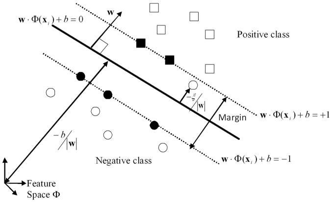
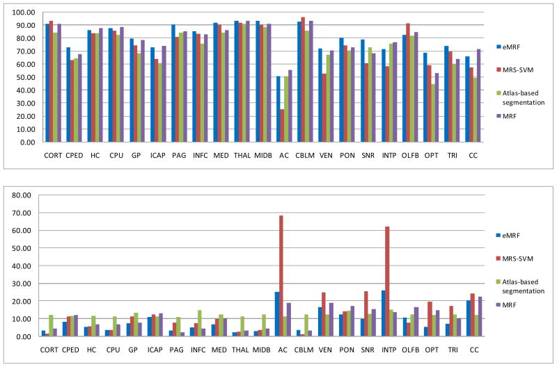
We introduce an automated segmentation method, extended Markov random field (eMRF), to classify 21 neuroanatomical structures of mouse brain based on three dimensional (3D) magnetic resonance images (MRI). The image data are multispectral: T2-weighted, proton density-weighted, diffusion x, y and z weighted. Earlier research (Ali, A.A., Dale, A.M., Badea, A., Johnson, G.A., 2005. Automated segmentation of neuroanatomical structures in multispectral MR microscopy of the mouse brain. NeuroImage 27 (2), 425-435) successfully explored the use of MRF for mouse brain segmentation. In this research, we study the use of information generated from support vector machine (SVM) to represent the probabilistic information. Since SVM in general has a stronger discriminative power than the Gaussian likelihood method and is able to handle nonlinear classification problems, integrating SVM into MRF improved the classification accuracy. The eMRF employs the posterior probability distribution obtained from SVM to generate a classification based on the MR intensity. Secondly, the eMRF introduces a new potential function based on location information. Third, to maximize the classification performance, the eMRF uses the contribution weights optimally determined for each of the three potential functions: observation, location and contextual functions, which are traditionally equally weighted. We use the voxel overlap percentage and volume difference percentage to evaluate the accuracy of eMRF segmentation and compare the algorithm with three other segmentation methods--mixed ratio sampling SVM (MRS-SVM), atlas-based segmentation and MRF. Validation using classification accuracy indices between automatically segmented and manually traced data shows that eMRF outperforms other methods.
Figures

References
-
- Ali AA, Dale AM, Badea A, Johnson GA. Automated segmentation of neuroanatomical structures in multispectral MR microscopy of the mouse brain. NeuroImage. 2005;27 (2):425–435. - PubMed
-
- Amato U, Larobina M, Antoniadis A, Alfano B. Segmentation of magnetic resonance images through discriminant analysis. Journal of Neuroscience Methods. 2003;131:65–74. - PubMed
-
- Andersen AH, Zhang Z, Avison MJ, Gash DM. Automated segmentation of multispectral brain MR images. Journal of Neuroscience Methods. 2002;122:13–23. - PubMed
-
- Ashburner J, Friston K. Multimodal image coregistration and partitioning – a unified framework. NeuroImage. 1997;6:209–217. - PubMed
-
- Bae MH, Wu T, Pan R. Mix-Ratio Sampling: Classifying Multiclass Imbalanced Mouse Brain Images Using Support Vector Machine. 2008. Technical Report available at http://swag.eas.asu.edu/vcie/
Publication types
MeSH terms
Grants and funding
LinkOut - more resources
Full Text Sources
Other Literature Sources
Medical
Miscellaneous
