. 2009 May;77(5):2065-75.
doi: 10.1128/IAI.01204-08. Epub 2009 Feb 23.

Role of LecA and LecB lectins in Pseudomonas aeruginosa-induced lung injury and effect of carbohydrate ligands

Affiliations

Role of LecA and LecB lectins in Pseudomonas aeruginosa-induced lung injury and effect of carbohydrate ligands

Chanez Chemani et al. Infect Immun. 2009 May.

Abstract

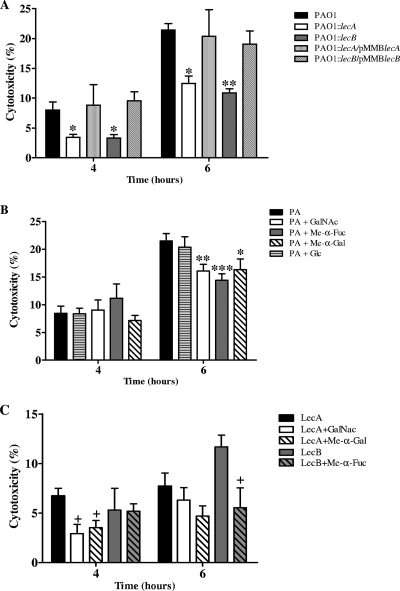
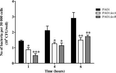
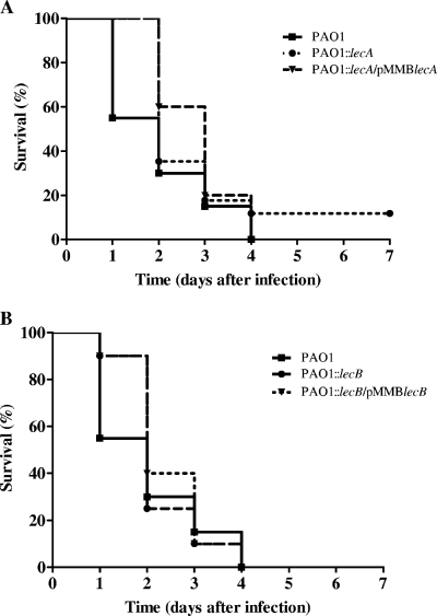
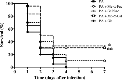
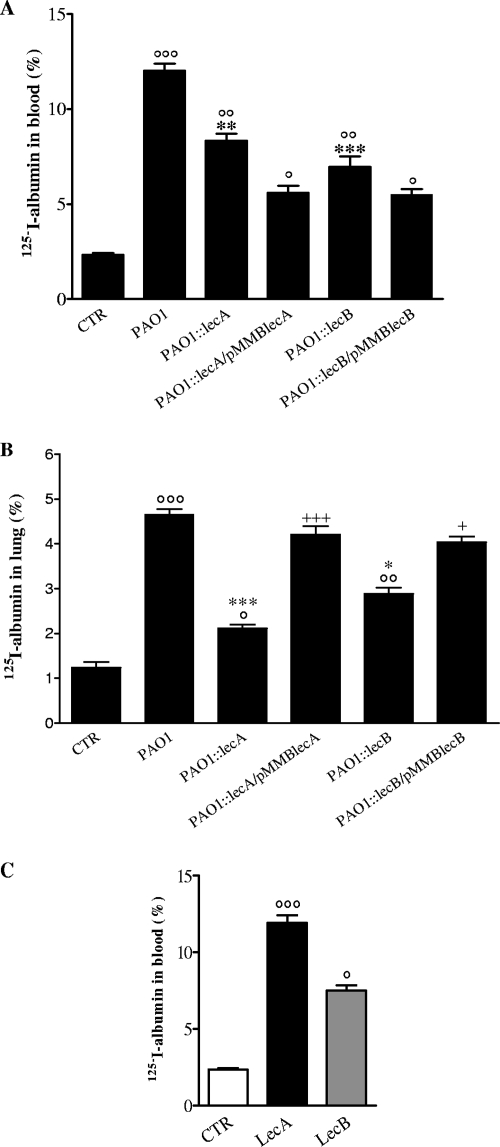
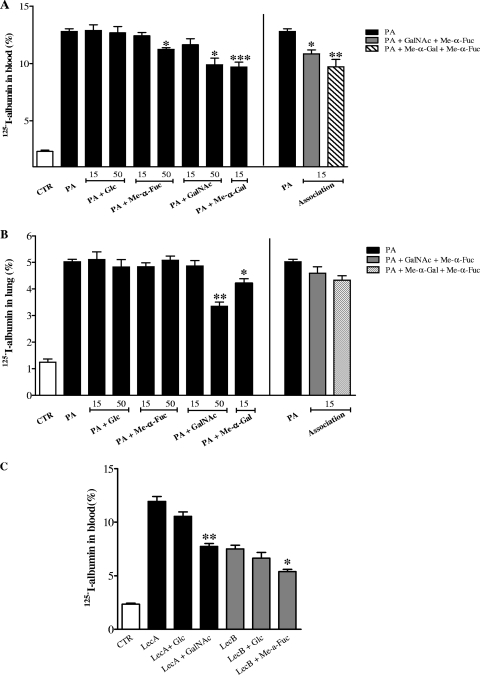
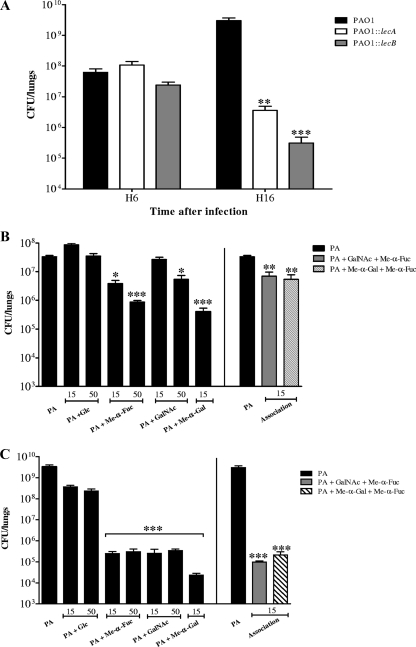
Pseudomonas aeruginosa is a frequently encountered pathogen that is involved in acute and chronic lung infections. Lectin-mediated bacterium-cell recognition and adhesion are critical steps in initiating P. aeruginosa pathogenesis. This study was designed to evaluate the contributions of LecA and LecB to the pathogenesis of P. aeruginosa-mediated acute lung injury. Using an in vitro model with A549 cells and an experimental in vivo murine model of acute lung injury, we compared the parental strain to lecA and lecB mutants. The effects of both LecA- and Lec B-specific lectin-inhibiting carbohydrates (alpha-methyl-galactoside and alpha-methyl-fucoside, respectively) were evaluated. In vitro, the parental strain was associated with increased cytotoxicity and adhesion on A549 cells compared to the lecA and lecB mutants. In vivo, the P. aeruginosa-induced increase in alveolar barrier permeability was reduced with both mutants. The bacterial burden and dissemination were decreased for both mutants compared with the parental strain. Coadministration of specific lectin inhibitors markedly reduced lung injury and mortality. Our results demonstrate that there is a relationship between lectins and the pathogenicity of P. aeruginosa. Inhibition of the lectins by specific carbohydrates may provide new therapeutic perspectives.


Figures

FIG. 1.
FIG. 1.
(A) PCR verification of lecA and lecB gene interruption using external lecA and lecB oligonucleotide pairs with which PCR amplification of the corresponding DNA region leads in the wild-type strain to 736- and 836-nucleotide DNA fragments for lecA and lecB, respectively. Lanes 1 and 4, parental PAO1 strain; lanes 2 and 5, PAO1lecA::Tcr and PAO1lecB::Tcr mutants, respectively; lanes 3 and 6, reference PAO1 strain. (B) Western blot analysis of LecA production in the parental strain P. aeruginosa PAO1 (lanes 1 to 3), PAO1lecA::Tcr insertional mutants (lanes 4 to 5), and the PAO1::lecA/pMMBlecA strain (lane 6) with a polyclonal antibody directed against the LecA protein (lane 7). The numbers above the Western blot indicate the growth points at which the production was checked (1, 2, and 3 indicate aliquots obtained during the exponential, early stationary, and late stationary phases, respectively). (C) Growth curves for the parental strain P. aeruginosa PAO1 and insertional mutants PAO1lecA::Tcr and PAO1lecB::Tcr. nt, nucleotides.
FIG. 2.
FIG. 2.
(A) Cytotoxicity of P. aeruginosa strains with lung epithelial cells. A549 cells (2 × 10⁴ cells) were cocultured with each strain at a multiplicity of infection of 250. (B and C) Effects of carbohydrates on the cytotoxicity of P. aeruginosa (PA) (B) and its lectins (C). A549 cells (2 × 10⁴ cells) were cocultured with P. aeruginosa PAO1 (parental strain) at a multiplicity of infection of 250 or with each lectin in the presence of Glc, GalNAc, Me-α-Fuc, or Me-α-Gal. The carbohydrates were added at a concentration of 15 mM. The cytotoxicity with lung epithelial cells was evaluated by measuring the release of LDH at 4 and 6 h. The values are the averages of three assays for the means (bars) and standard deviations (error bars). *, P < 0.05 for a comparison with PAO1; **, P < 0.01 for a comparison with PAO1; ***, P < 0.001 for a comparison with PAO1; +, P < 0.05 for a comparison with the corresponding lectin.
FIG. 3.
FIG. 3.
Relative attachment of P. aeruginosa strains to A549 cells: numbers of P. aeruginosa PAO1 (parental strain), PAO1::lecA (lecA mutant), and PAO1::lecB (lecB mutant) bacteria adhering to A549 cells. An inoculum containing 108 CFU/ml was used. Bacteria were allowed to attach for 1, 4, and 6 h. The values are the means (bars) and standard deviations (error bars) of five independent experiments. *, P < 0.05; **, P < 0.01; ***, P < 0.001 (for a comparison with the PAO1 group).
FIG. 4.
FIG. 4.
Survival study. An inoculum containing 5 × 10⁷ CFU was instilled into the lungs, and then survival was monitored for 7 days for 20 mice per group. (A) Survival of mice after instillation of PAO1::lecA compared with the survival after instillation of PAO1 (parental strain) or PAO1::lecA/pMMBlecA (complemented lecA mutant). (B) Survival of mice after instillation of PAO1::lecB (lecB mutant) compared with the survival after instillation of PAO1 (parental strain) or PAO1::lecB/pMMBlecB (complemented lecB mutant).
FIG. 5.
FIG. 5.
Effect of carbohydrates on mortality in mice. Mice were inoculated intratracheally with a suspension containing 5 × 10⁷ CFU of P. aeruginosa PAO1 (parental strain) in the presence and absence of Glc, GalNAc, Me-α-Gal, or Me-α-Fuc, and then survival was monitored for 7 days. The carbohydrates were used at a concentration of 15 mM (20 mice per group). *, P < 0.05 for a comparison with the PAO1 group; **, P < 0.001 for a comparison with the PAO1 group. PA, P. aeruginosa.
FIG. 6.
FIG. 6.
Alveolar capillary barrier permeability after intratracheal instillation of P. aeruginosa PAO1 (parental strain), PAO1::lecA (lecA mutant), PAO1::lecB (lecB mutant), and P. aeruginosa lectins (LecA and LecB). (A and C) Efflux of the protein tracer 125I-albumin from the lungs into the blood 6 h after infection. (B) Efflux of the protein tracer 125I-albumin from the blood into the lungs 16 h after intraperitoneal injection. The data are means (bars) and standards errors (error bars) for 10 mice per group. °, P < 0.05 for a comparison with the control; °°, P < 0.01 for a comparison with the control; °°°, P < 0.001 for a comparison with the control; *, P < 0.05 for a comparison with PAO1; **, P < 0.01 for a comparison with PAO1; ***, P < 0.001 for a comparison with PAO1; +, P < 0.05 for a comparison with the lecB mutant or lecA mutant; +++, P < 0.001 for a comparison with the lecB mutant or lecA mutant. CTR, control (no bacteria or no lectin).
FIG. 7.
FIG. 7.
Effect of carbohydrates on lung injury. Lung injury was evaluated by examining alveolar capillary barrier permeability after intratracheal instillation of P. aeruginosa PAO1 (parental strain) and its lectins alone or in combination with Glc, GalNAc, Me-α-Gal, or Me-α-Fuc in mice. (A and C) Efflux of the protein tracer 125I-albumin from the lungs into the blood 6 h after infection. (B) Efflux of the protein tracer 125I-albumin from the blood into the lungs 16 h after intraperitoneal injection. The data are means (bars) and standards errors (error bars). The carbohydrates were used at concentrations of 15 and 50 mM, and there were 10 mice per group. *, P < 0.05 for a comparison with PAO1; **, P < 0.01 for a comparison with PAO1; ***, P < 0.001 for a comparison with PAO1. CTR, control (no bacteria or no lectin); PA, P. aeruginosa.
FIG. 8.
FIG. 8.
Lung bacterial clearance. Mice were infected with a suspension containing 5 × 10⁶ CFU of P. aeruginosa PAO1 (parental strain), PAO1::lecA (lecA mutant), or PAO1::lecB (lecB mutant). The number of viable bacteria remaining in the infected lungs was counted 6 h (H6) and 16 h (H16) after instillation (A). The effect of carbohydrates on lung bacterial clearance was evaluated after intratracheal instillation of 5 × 10⁷ CFU of P. aeruginosa PAO1 mixed with Glc, GalNAc, Me-α-Gal, or Me-α-Fuc at 6 h (B) and 16 h (C). Carbohydrates were used at concentrations of 15 and 50 mM, and there were 10 mice per group. The data are means (bars) and standard errors (error bars). *, P < 0.05 for a comparison with PAO1; **, P < 0.01 for a comparison with PAO1; ***, P < 0.001 for a comparison with PAO1. PA, P. aeruginosa.

References

    1. Ader, F., B. R. Le, K. Faure, P. Gosset, O. Epaulard, B. Toussaint, B. Polack, E. Nowak, N. B. Viget, E. Kipnis, and B. P. Guery. 2005. Alveolar response to Pseudomonas aeruginosa: role of the type III secretion system. Infect. Immun. 734263-4271. - PMC - PubMed
    1. Bajolet-Laudinat, O., S. Girod-de Bentzmann, J. M. Tournier, C. Madoulet, M. C. Plotkowski, C. Chippaux, and E. Puchelle. 1994. Cytotoxicity of Pseudomonas aeruginosa internal lectin PA-I to respiratory epithelial cells in primary culture. Infect. Immun. 624481-4487. - PMC - PubMed
    1. Bryan, R., M. Feldman, S. C. Jawetz, S. Rajan, E. DiMango, H. B. Tang, L. Scheffler, D. P. Speert, and A. Prince. 1999. The effects of aerosolized dextran in a mouse model of Pseudomonas aeruginosa pulmonary infection. J. Infect. Dis. 1791449-1458. - PubMed
    1. Chen, C. P., S. C. Song, N. Gilboa-Garber, K. S. Chang, and A. M. Wu. 1998. Studies on the binding site of the galactose-specific agglutinin PA-IL from Pseudomonas aeruginosa. Glycobiology 87-16. - PubMed
    1. Cioci, G., E. P. Mitchell, C. Gautier, M. Wimmerova, D. Sudakevitz, S. Perez, N. Gilboa-Garber, and A. Imberty. 2003. Structural basis of calcium and galactose recognition by the lectin PA-IL of Pseudomonas aeruginosa. FEBS Lett. 555297-301. - PubMed

Publication types

MeSH terms

LinkOut - more resources