. 2009 May;77(5):1807-16.
doi: 10.1128/IAI.01162-08. Epub 2009 Feb 23.

Yersinia pestis IS1541 transposition provides for escape from plague immunity

Affiliations

Yersinia pestis IS1541 transposition provides for escape from plague immunity

Claire A Cornelius et al. Infect Immun. 2009 May.

Abstract

Yersinia pestis is perhaps the most feared infectious agent due to its ability to cause epidemic outbreaks of plague disease in animals and humans with high mortality. Plague infections elicit strong humoral immune responses against the capsular antigen (fraction 1 [F1]) of Y. pestis, and F1-specific antibodies provide protective immunity. Here we asked whether Y. pestis generates mutations that enable bacterial escape from protective immunity and isolated a variant with an IS1541 insertion in caf1A encoding the F1 outer membrane usher. The caf1A::IS1541 insertion prevented assembly of F1 pili and provided escape from plague immunity via F1-specific antibodies without a reduction in virulence in mouse models of bubonic or pneumonic plague. F1-specific antibodies interfere with Y. pestis type III transport of effector proteins into host cells, an inhibitory effect that was overcome by the caf1A::IS1541 insertion. These findings suggest a model in which IS1541 insertion into caf1A provides for reversible changes in envelope structure, enabling Y. pestis to escape from adaptive immune responses and plague immunity.


Figures
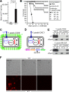
FIG. 1.
FIG. 1.
Isolation of Y. pestis escape variants. (A) Mice were immunized with recombinant capsular antigen (rF1) or a PBS control, and serum IgG antibody titers were determined by ELISA. (B) rF1-immunized mice (cohorts of 10 animals) were challenged with increasing doses of wild-type strain Y. pestis CO92, and their survival was monitored. (C) Diagram showing Y. pestis CO92 and its virulence plasmids pCD1 (providing LcrV and type III secretion [blue]) and pFra (with the caf1R gene and the caf1M-caf1A-caf1 operon for biogenesis of F1 pili [green]), as well as the chromosome with a high-pathogenicity island-pigmentation segment (pgm) (red) and IS1541 elements (arrowheads). (D) Insertion of IS1541 into caf1A abrogates F1 secretion and capsule assembly. (E) Cultures of Y. pestis CO92, KIM D27 (Δpgm), and isogenic variants with a caf1 deletion (ΔF1) or caf1A::IS1541 (CAC1 and CAC2) were centrifuged, and proteins in the culture supernatant (S) and bacterial pellet (P) were analyzed by immunoblotting with F1-specific antibody (αF1) and RNA polymerase-specific antibody (αRpoA). (F) Y. pestis strains were analyzed by differential interference contrast (DIC) or fluorescence microscopy with F1-specific antibody. s.c., subcutaneous; WT, wild type.
FIG. 2.
FIG. 2.
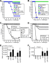
Y. pestis with caf1A::IS1541 is fully virulent. (A) Mice were challenged by subcutaneous (s.c.) injection with the indicated numbers of CFU of Y. pestis CO92 (wild type) or CAC1 (caf1A::IS1541) to precipitate bubonic plague, and their survival was monitored. (B) Strains were inoculated intranasally (i.n.) into mice to precipitate pneumonic plague. (C) Y. pestis strains were inoculated intranasally, and lung tissue replication and dissemination of plague bacteria into the spleen were measured on three consecutive days. (D) Lung tissue from animals infected as described above for panel C was fixed, thin sectioned, stained with hematoxylin-eosin, and viewed by microscopy.
FIG. 3.
FIG. 3.
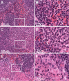
Spleens from naïve BALB/c mice (A and B) and BALB/c mice infected via subcutaneous injection with 20 CFU of Y. pestis CO92 (C and D) or Y. pestis CAC1 (E and F) were removed during necropsy on day 7 following the infectious challenge. Tissues were fixed, embedded in paraffin, thin sectioned, stained with hematoxylin-eosin, and viewed by light microscopy at a magnification of ×40. Captured images (A, C, and E) were enlarged fivefold to reveal loss of tissue architecture, necrotic polymorphonuclear leukocytes, massive cellular debris, and edema in plague-infected spleens (D and F) compared to uninfected tissue (B).
FIG. 4.
FIG. 4.
Y. pestis with caf1A::IS1541 escapes plague protective immunity. (A) Mice were immunized with the nonpigmented (Δpgm) strain KIM D27 or mock treated (PBS) and infected by subcutaneous (s.c.) injection with Y. pestis CO92 or CAC1 (caf1A::IS1541), and their survival after bubonic plague challenge was monitored. (B) Mice were immunized as described above for panel A and infected by intranasal (i.n.) instillation, and their survival after pneumonic plague challenge was monitored. (C) Mice were immunized with a mock control (PBS) or with purified subunit vaccines (rLcrV, rV10, rF1, rLcrV plus F1, and rV10 plus F1) and challenged by subcutaneous injection with Y. pestis CAC1, and their survival was monitored. (D) Mice immunized as described above for panel C were challenged by intranasal instillation with Y. pestis CAC1, and their survival was monitored. Sera from mice immunized with subunit vaccines as described above for panels C and D were examined by ELISA for LcrV-specific (E) and F1-specific (F) IgG antibody titers.
FIG. 5.
FIG. 5.
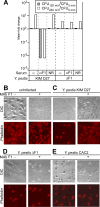
F1 antibodies block Y. pestis type III injection, which is overcome by caf1A::IS1541. (A) Human anticoagulated blood was incubated for 120 or 240 min with 105 CFU Y. pestis KIM D27 or the isogenic ΔF1 mutant strain in the absence (−) or presence of mouse anti-F1 (αF1) or a nonreactive serum control (NR). Mean changes in bacterial load were recorded by plating aliquots on agar and incubating the preparations for colony formation (CFU). (B to E) HeLa tissue culture cells were viewed by differential interference contrast (DIC) microscopy, or their actin cables were stained with rhodamine-phalloidin and viewed by fluorescence microscopy. Tissue culture cells were either not infected (B) or were infected with plague strains in the presence or absence of 500 μg F1-specific MAb F1-04-A-G1 (MAb F1) using Y. pestis KIM D27 (C), ΔF1 (caf1 deletion) (D), and CAC2 (caf1A::IS1541) (E).

References

    1. Anderson, G. W., Jr., P. L. Worsham, C. R. Bolt, G. P. Andrews, S. L. Welkos, A. M. Friedlander, and J. P. Burans. 1997. Protection of mice from fatal bubonic and pneumonic plague by passive immunization with monoclonal antibodies against the F1 protein of Yersinia pestis. Am. J. Trop. Med. Hyg. 56471-473. - PubMed
    1. Andrews, G. P., D. G. Heath, G. W. Anderson, Jr., S. L. Welkos, and A. M. Friedlander. 1996. Fraction 1 capsular antigen (F1) purification from Yersinia pestis CO92 and from an Escherichia coli recombinant strain and efficacy against lethal plague challenge. Infect. Immun. 642180-2187. - PMC - PubMed
    1. Anisimov, A., L. Lindler, and G. Pier. 2004. Intraspecific diversity of Yersinia pestis. Clin. Microbiol. Rev. 17434-464. - PMC - PubMed
    1. Baker, E. E., H. Sommer, L. W. Foster, E. Meyer, and K. F. Meyer. 1952. Studies on immunization against plague. I. The isolation and characterization of the soluble antigen of Pasteurella pestis. J. Immunol. 68131-145. - PubMed
    1. Blomfield, I. C., V. Vaughn, R. F. Rest, and B. I. Eisenstein. 1991. Allelic exchange in Escherichia coli using the Bacillus subtilis sacB gene and a temperature-sensitive pSC101 replicon. Mol. Microbiol. 51447-1457. - PubMed

Publication types

MeSH terms

Associated data