. 2009 Apr;29(8):2053-67.
doi: 10.1128/MCB.01179-08. Epub 2009 Feb 23.

Peroxisome proliferator-activated receptor gamma activation restores islet function in diabetic mice through reduction of endoplasmic reticulum stress and maintenance of euchromatin structure

Affiliations

Peroxisome proliferator-activated receptor gamma activation restores islet function in diabetic mice through reduction of endoplasmic reticulum stress and maintenance of euchromatin structure

Carmella Evans-Molina et al. Mol Cell Biol. 2009 Apr.

Abstract

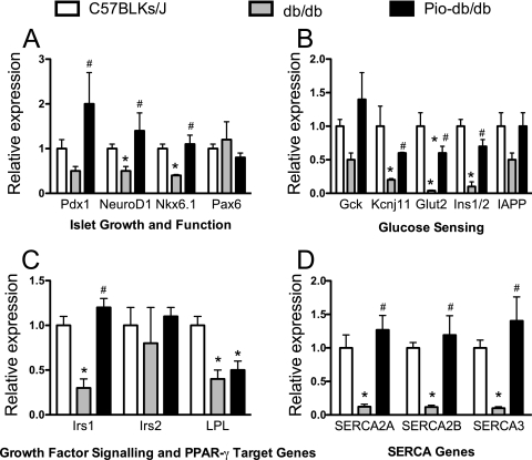
The nuclear receptor peroxisome proliferator-activated receptor gamma (PPAR-gamma) is an important target in diabetes therapy, but its direct role, if any, in the restoration of islet function has remained controversial. To identify potential molecular mechanisms of PPAR-gamma in the islet, we treated diabetic or glucose-intolerant mice with the PPAR-gamma agonist pioglitazone or with a control. Treated mice exhibited significantly improved glycemic control, corresponding to increased serum insulin and enhanced glucose-stimulated insulin release and Ca(2+) responses from isolated islets in vitro. This improved islet function was at least partially attributed to significant upregulation of the islet genes Irs1, SERCA, Ins1/2, and Glut2 in treated animals. The restoration of the Ins1/2 and Glut2 genes corresponded to a two- to threefold increase in the euchromatin marker histone H3 dimethyl-Lys4 at their respective promoters and was coincident with increased nuclear occupancy of the islet methyltransferase Set7/9. Analysis of diabetic islets in vitro suggested that these effects resulting from the presence of the PPAR-gamma agonist may be secondary to improvements in endoplasmic reticulum stress. Consistent with this possibility, incubation of thapsigargin-treated INS-1 beta cells with the PPAR-gamma agonist resulted in the reduction of endoplasmic reticulum stress and restoration of Pdx1 protein levels and Set7/9 nuclear occupancy. We conclude that PPAR-gamma agonists exert a direct effect in diabetic islets to reduce endoplasmic reticulum stress and enhance Pdx1 levels, leading to favorable alterations of the islet gene chromatin architecture.


Figures

FIG. 1.
FIG. 1.
Pioglitazone treatment results in greater weight gain but improved serum lipids in db/db mice. Male C57BLKS/J-db/db mice were treated with either vehicle (db/db) or pioglitazone (Pio-db/db) for 6 weeks and compared to age- and sex-matched lean C57BLKS/J mice. Following the treatment period, mice were evaluated for body weight (A), serum total cholesterol (B), serum triglycerides (C), and serum free fatty acids (D). Data represent the means ± standard errors of the results obtained with 12 mice per group. *, significantly (P < 0.05) different compared to C57BLKS/J mouse results; #, significantly (P < 0.05) different compared to db/db mouse results.
FIG. 2.
FIG. 2.
Pioglitazone treatment improves glycemic control and insulin levels in db/db mice. Male db/db mice were treated with either vehicle (db/db) or pioglitazone (Pio-db/db) for 6 weeks and compared to age- and sex-matched lean C57BLKS/J mice. (A) Results of random blood glucose tests at the end of the 6-week treatment period; (B) results of intraperitoneal glucose tolerance tests at the end of the 6-week treatment period. Pio-db/db animals exhibited significantly improved glucose tolerance compared to db/db animals (P < 0.001 for the comparison by two-way ANOVA). (C) Random serum insulin levels at the end of the 6-week treatment period; (D) results of insulin tolerance tests. No differences in insulin tolerance were observed between the db/db and Pio-db/db groups, whereas both groups had significantly impaired insulin tolerance compared to the C57BLKS/J mouse group (P < 0.001 for the comparison by two-way ANOVA). Data represent the means ± standard errors of the results obtained with at least 12 animals per group. *, significantly (P < 0.05) different compared to C57BLKS/J mice; #, significantly (P < 0.05) different compared to db/db mice.
FIG. 3.
FIG. 3.
Islet architecture and insulin staining. Pancreata from male db/db mice, treated with either vehicle (db/db) or pioglitazone (Pio-db/db) for 6 weeks, were fixed and stained and compared to pancreata from age- and sex-matched lean C57BLKS/J mice. (A, B, and C) Hematoxylin and eosin staining of pancreatic sections from C57BLKS/J, db/db, and Pio-db/db mice; (D, E, and F) immunofluorescence staining of islets from C57BLKS/J, db/db, and Pio-db/db mice for insulin (red) and glucagon (green). Nuclei were counterstained with Hoechst dye (blue). The figure shows representative islets from three pancreata analyzed per group of mice.
FIG. 4.
FIG. 4.
Pioglitazone treatment improves islet function in db/db mice. Islets from male db/db mice, treated with either vehicle (db/db) or pioglitazone (Pio-db/db) for 6 weeks, were compared to islets from age-matched lean C57BLKS/J mice. (A) Results of GSCa studies of isolated islets. The panel shows the continuous fura-2 AM fluorescence ratio (340/380 nm) as glucose in the incubation chamber was increased from 3 mM to 28 mM. Data represent the means ± standard errors of the results obtained with at least 30 islets from 12 different animals per group. (B) Data from panel A were used to calculate a GSCa index, which represents the fura-2 AM fluorescence ratio at 28 mM glucose divided by the ratio at 3 mM glucose. (C) The insulin content of islets used for static release assays was measured by ELISA after acid extraction. (D) Islets were incubated in 3 and 28 mM glucose for 1 h, and insulin secretion into the supernatant was measured by ELISA. Data represent the means of the results of at least three independent experiments performed using 50 islets per group. *, significantly (P < 0.05) different compared to C57BLKS/J mice; #, significantly (P < 0.05) different compared to db/db mice.
FIG. 5.
FIG. 5.
Pioglitazone treatment improves islet calcium oscillations. Islets from male db/db mice, treated with either vehicle (db/db) or pioglitazone (Pio-db/db) for 6 weeks, were harvested and compared to islets from age-matched lean C57BLKS/J mice. (A to C) Three representative calcium oscillations at an 11 mM glucose concentration from islets isolated from C57BLKS/J mice (A), db/db mice (B), and Pio-db/db mice (C). (D) Percentages of islets exhibiting endogenous calcium oscillations; (E) amplitude of calcium oscillations; (F) period of oscillations. Data in panels D to F represent results obtained from analysis of at least 30 islets from 12 different animals per group. *, statistically (P < 0.05) different compared to C57BLKS/J mice; #, statistically (P < 0.05) different compared to db/db mice.
FIG. 6.
FIG. 6.
Pioglitazone treatment improves islet function in mice fed an HFD. Male C57BLKS/J mice were fed an HFD (42% of calories from fat) and gavaged with either vehicle (HFD) or pioglitazone (HFD+Pio) for 4 weeks. Results were compared to those obtained with age- and sex-matched C57BLKS/J mice fed a regular chow diet (17% of calories from fat [Chow]). (A) Body weight at end of study; (B) results of intraperitoneal glucose tolerance tests at the end of study. Pio + HFD animals exhibited significantly improved glucose tolerance compared to HFD animals (P < 0.05 for the comparison by two-way ANOVA). (C) Results of analysis of serum insulin levels at times 0 and 30 min during the glucose tolerance test represented in panel B. (D) Islets were incubated in 3 and 28 mM glucose for 1 h, and insulin secretion into the supernatant was measured by ELISA. Data represent the means of the results of at least three independent experiments performed using 50 islets per group. The results shown in panels C and D were analyzed by one-way ANOVA. #, significantly (P < 0.05) different compared to Chow mice; *, significantly (P < 0.05) different compared to HFD mice.
FIG. 7.
FIG. 7.
Pioglitazone treatment improves the expression of key β-cell genes. Islets from age- and sex-matched male C57BLKS/J and db/db mice, treated with either vehicle (db/db) or pioglitazone (Pio-db/db) for 6 weeks, were harvested and subjected to real-time RT-PCR for analysis of islet growth and function genes (A), glucose-sensing genes (B), growth factor-signaling genes (C), and SERCA genes (D). Data represent the means ± standard errors of the results of at least three different biologic replicate experiments. Data were analyzed by one-way ANOVA. *, statistically (P < 0.05) different compared to C57BLKS/J mice; #, statistically (P < 0.05) different compared to db/db mice.
FIG. 8.
FIG. 8.
Pioglitazone treatment enhances euchromatin markers at the Ins1/2 and Glut2 promoters. Islets from male db/db mice, treated with either vehicle (db/db) or pioglitazone (Pio-db/db) for 6 weeks, were harvested and subjected to ChIP analysis as detailed in Materials and Methods. Real-time PCR was used to quantitate recovery of the proximal Ins1/2 and Glut2 promoters, and results are expressed as percent recovery of the gene fragment relative to input chromatin. (A) ChIP using normal rabbit serum (NRS) or antibody to acetylated H4 (Ac H4), followed by analysis of recovered Ins1/2 promoter fragments; (B) ChIP using NRS or antibody to acetylated H3 (Ac H4) followed by analysis of recovered Ins1/2 promoter fragments; (C) ChIP using NRS or antibody to H3-dimethyl-Lys4, followed by analysis of recovered Ins1/2 promoter fragments; (D) ChIP using NRS or antibody to H3-dimethyl-Lys4, followed by analysis of recovered Glut2 promoter fragments. Results represent the means ± standard errors of the results of three independent ChIP experiments. Results were analyzed using either a t test or one-way ANOVA (C). *, results were significantly (P < 0.05) different for the comparison shown.
FIG. 9.
FIG. 9.
Expression patterns of Pdx-1 and the methyltransferase Set7/9. Islets and pancreata from male db/db mice, treated with either vehicle (db/db) or pioglitazone (Pio-db/db), were harvested and subjected to real-time RT-PCR, immunoblotting, or immunohistochemistry. (A) Results of real-time RT-PCR analysis for Setd7 mRNA by the use of RNA from isolated islets. Data represent the means ± standard errors of the results obtained from two independent islet isolation experiments using six animals in each experiment. (B) Results of immunoblot analysis for Set7/9 (upper panel), Pdx-1 (lower panel), or GAPDH (both panels) obtained using total protein from isolated islets. Data are from pooled-islet experiments performed using six different animals per group. Data from a second islet pool were similar. (C to H) Pancreata from db/db and Pio-db/db animals were peroxidase stained for Set7/9 (C and D) and Pdx-1 (E and F) and counterstained with hematoxylin or stained for both Set7/9 (green) and Pdx-1 (red) and visualized by immunofluorescence (G and H). Nuclei were counterstained using Hoechst dye in (G and H). Data in panels C to H show representative islets from among three pancreata analyzed per group of mice.
FIG. 10.
FIG. 10.
PPAR-γ activation in vitro improves glucose-stimulated calcium response in db/db islets. Islets from 8-week-old male db/db mice were incubated for 24 h with 10 μM pioglitazone (Pio-db/db) or vehicle control (db/db). (A) Results of GSCa studies of isolated islets. The panel shows the continuous fura-2 AM fluorescence ratio (340/380 nm) changes as glucose in the incubation chamber was increased from 3 mM to 28 mM. Data represent the means ± standard errors of the results for at least 10 islets per group. (B) Data from panel A were used to calculate a GSCa index, which represents the fura-2 AM fluorescence ratio under conditions of 28 mM glucose divided by the ratio under conditions of 3 mM glucose. (C) Islets were incubated in 3 and 28 mM glucose for 1 h, and insulin secretion into the supernatant was measured by ELISA. Data represent the means of the results of at least three independent experiments performed using 50 islets per group. Results were analyzed using a t test. *, statistically different (P < 0.05) compared to db/db islets treated with vehicle control.
FIG. 11.
FIG. 11.
PPAR-γ activation in vitro upregulates SERCA genes and reduces ER stress in db/db islets. Islets from 9- to 10-week-old db/db mice, treated with either vehicle (db/db) or 10 μM pioglitazone (db/db + Pio) for 24 h, were subjected to real-time RT-PCR for analysis of SERCA genes (A) and ER stress markers (B). Data represent the means ± standard errors of the results of five biologic replicate experiments. Results were analyzed by one-way ANOVA. *, significantly (P < 0.05) different compared to db/db mouse group results.
FIG. 12.
FIG. 12.
PPAR-γ activation in vitro reduces ER stress in INS-1 (832/13) β cells. (A) INS-1 β cells were incubated with vehicle (DMSO), 1 μM thapsigargin (Thap), or 1 μM thapsigargin plus 10 μM pioglitazone (Thap + Pio) for 6 h and then subjected to immunoblot analysis for the proteins indicated. (B to D) Quantitation of the immunoblots shown in panel A. Data represent the means ± standard errors of the results of three independent biological replicate experiments. Data were analyzed by one-way ANOVA, and statistical significance (P < 0.05) was determined for the data in panel D for the comparison of Thap versus DMSO and of Thap + Pio versus Thap.
FIG. 13.
FIG. 13.
PPAR-γ activation in vitro promotes nuclear recovery of Set7/9 in INS-1 (832/13) β cells. INS-1 β cells were incubated with vehicle (DMSO) (A and B), 1 μM thapsigargin (Thap) (C and D), or 1 μM thapsigargin plus 10 μM pioglitazone (Thap + Pio) (E and F) for 6 h and were then subjected to fixation and immunostaining for Set7/9 or DAPI (to visualize nuclei), as indicated at the top. (G) The relative ratios of nuclear intensity to cytoplasmic intensity for Set7/9 were calculated for a minimum of 10 cells per set of conditions. Data in panel G were analyzed by one-way ANOVA, and statistical significance (P < 0.05) was obtained for the comparision of Thap versus DMSO and of Thap+Pio versus Thap.
FIG. 14.
FIG. 14.
Inducible deletion of Pdx1 in pancreatic islets causes a nuclear-to-cytoplasmic shift in Set7/9. Pdx1tTA/tTA; TgPdx1 mice (20) were treated with vehicle for 7 days as described in Materials and Methods, and pancreata were harvested and fixed for immunostaining for Pdx1, Set7/9, and DAPI, as indicated. (A to C) Pancreatic section from a representative control animal treated with vehicle (−DOXY); (D to F) pancreatic section from a representative animal treated with doxycycline (+DOXY).
FIG. 15.
FIG. 15.
Schematic diagram proposing a model for PPAR-γ action in the setting of diabetes or insulin resistance. The figure suggests two arms in the pathway through which PPAR-γ might improve glycemic control in the setting of diabetes and insulin resistance. The left arm proposes PPAR-γ action with respect to insulin-sensitive tissues such as muscle and adipose tissue, and the right arm proposes PPAR-γ action in the islet directly. See the text for details.

References

    1. Amrani, A., S. Durant, M. Throsby, J. Coulaud, M. Dardenne, and F. Homo-Delarche. 1998. Glucose homeostasis in the nonobese diabetic mouse at the prediabetic stage. Endocrinology 1391115-1124. - PubMed
    1. Brunham, L. R., J. K. Kruit, T. D. Pape, J. M. Timmins, A. Q. Reuwer, Z. Vasanji, B. J. Marsh, B. Rodrigues, J. D. Johnson, J. S. Parks, C. B. Verchere, and M. R. Hayden. 2007. Beta-cell ABCA1 influences insulin secretion, glucose homeostasis and response to thiazolidinedione treatment. Nat. Med. 13340-347. - PubMed
    1. Campbell, I. W., and S. Mariz. 2007. Beta-cell preservation with thiazolidinediones. Diabetes Res. Clin. Pract. 76163-176. - PubMed
    1. Chakrabarti, S. K., J. Francis, S. M. Ziesmann, J. C. Garmey, and R. G. Mirmira. 2003. Covalent histone modifications underlie the developmental regulation of insulin gene transcription in pancreatic beta cells. J. Biol. Chem. 27823617-23623. - PubMed
    1. Chakrabarti, S. K., J. C. James, and R. G. Mirmira. 2002. Quantitative assessment of gene targeting in vitro and in vivo by the pancreatic transcription factor, Pdx1. Importance of chromatin structure in directing promoter binding. J. Biol. Chem. 27713286-13293. - PubMed

Publication types

MeSH terms

LinkOut - more resources