. 2009 May 29;284(22):14838-48.
doi: 10.1074/jbc.M809542200. Epub 2009 Feb 23.

Placenta-specific methylation of the vitamin D 24-hydroxylase gene: implications for feedback autoregulation of active vitamin D levels at the fetomaternal interface

Affiliations

Placenta-specific methylation of the vitamin D 24-hydroxylase gene: implications for feedback autoregulation of active vitamin D levels at the fetomaternal interface

Boris Novakovic et al. J Biol Chem. .

Abstract

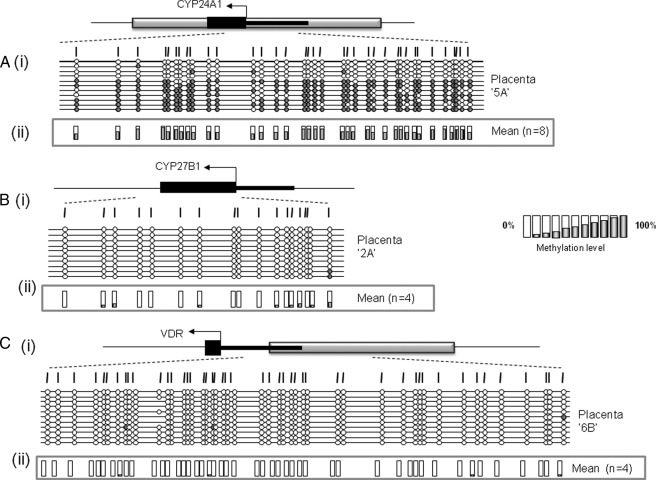
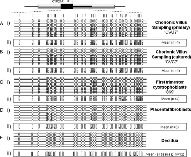
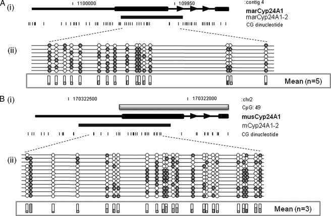
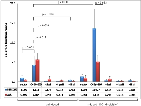
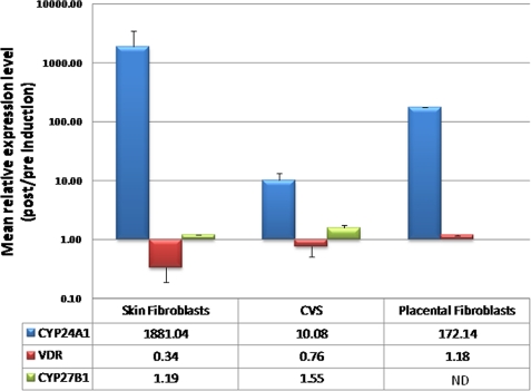
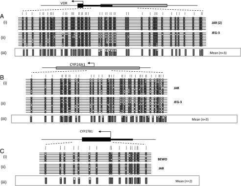
Plasma concentrations of biologically active vitamin D (1,25-(OH)(2)D) are tightly controlled via feedback regulation of renal 1alpha-hydroxylase (CYP27B1; positive) and 24-hydroxylase (CYP24A1; catabolic) enzymes. In pregnancy, this regulation is uncoupled, and 1,25-(OH)(2)D levels are significantly elevated, suggesting a role in pregnancy progression. Epigenetic regulation of CYP27B1 and CYP24A1 has previously been described in cell and animal models, and despite emerging evidence for a critical role of epigenetics in placentation generally, little is known about the regulation of enzymes modulating vitamin D homeostasis at the fetomaternal interface. In this study, we investigated the methylation status of genes regulating vitamin D bioavailability and activity in the placenta. No methylation of the VDR (vitamin D receptor) and CYP27B1 genes was found in any placental tissues. In contrast, the CYP24A1 gene is methylated in human placenta, purified cytotrophoblasts, and primary and cultured chorionic villus sampling tissue. No methylation was detected in any somatic human tissue tested. Methylation was also evident in marmoset and mouse placental tissue. All three genes were hypermethylated in choriocarcinoma cell lines, highlighting the role of vitamin D deregulation in this cancer. Gene expression analysis confirmed a reduced capacity for CYP24A1 induction with promoter methylation in primary cells and in vitro reporter analysis demonstrated that promoter methylation directly down-regulates basal promoter activity and abolishes vitamin D-mediated feedback activation. This study strongly suggests that epigenetic decoupling of vitamin D feedback catabolism plays an important role in maximizing active vitamin D bioavailability at the fetomaternal interface.


Figures

FIGURE 1.
FIGURE 1.
Methylation status of CYP24A1, CYP27B1, and VDR in human term placenta. Schematic representation of methylation data generated for regulatory regions of genes coding for 24-hydroxylase (A), 1α-hydroxylase (B), and the vitamin D receptor (C). i, gene structure showing the location of methylation assays. Gray box, CpG island; black boxes, exon regions; arrow, translational start site. ii, mean methylation levels seen at each CpG site for each type of sample tested. Numbers of each type of tissue are listed in parentheses. Between 8 and 12 individual clones were sequenced for each sample. Circles, CpG sites denoted by vertical dashes. Closed circles, methylation; open circles, lack of methylation. Missing circles indicate CpG sites for which no information was obtained.
FIGURE 2.
FIGURE 2.
Methylation of CYP24A1 in different human placental tissues and cell types. Representative methylation data generated for the CYP24A1-2 assay in cytokeratin-7 positive cytotrophoblasts (A), uncultured CVS (B), cultured CVS (C), full term placental fibroblasts (D), and whole blood (E). i, between 8 and 12 individual clones were sequenced for each sample. Circles, CpG sites denoted by vertical dashes. Closed circles, methylation; open circles, lack of methylation. Missing circles indicate CpG sites for which no information was obtained. Gray boxes, to GpG island locations; arrows, start site of translation within exon 1 (black line). ii, mean methylation levels seen at each CpG site for each type of sample tested. Numbers of each type of tissue are listed in parentheses.
FIGURE 3.
FIGURE 3.
Variable levels of CYP24A1 methylation in different mammalian full term placentas. Representative methylation data were generated for homologous CYP24A1 promoter region in marmoset (A) and mouse (B). i, gray boxes, GpG island locations; arrows, orientation of transcription. Black boxes, assay location. ii, eight individual clones were sequenced for each sample. Circles, CpG sites denoted by black dashes. Closed circles, methylation; open circles, lack of methylation. Shaded bars, mean methylation levels seen at each CpG site for each type of sample tested.
FIGURE 4.
FIGURE 4.
CYP24A1 promoter activity in human trophoblast cell lines. SV40-transformed cytotrophoblast (HIPEC-65) or choriocarcinoma (JAR) cells were transfected (n = 8 for each cell line) with a promoterless luciferase reporter (vector) with or without the cloned CYP24A1 gene promoter (–548/+209 relative to the start site of transcription). This region has previously been demonstrated to contain vitamin D-responsive elements (47). Basal promoter activity was detected in both cell lines and was abolished with in vitro methylation of the promoter prior to transfection (Sss1, HpaII, and HhaI methylases). 1,25-(OH)2D (active vitamin D) induction of the promoter was observed only in HIPEC-65 cells (JAR cells are predicted to lack the vitamin D receptor, as evidenced by complete methylation of the associated CpG island), but this induction was attenuated with prior methylation of the promoter region. Luminescence values were normalized to Renilla luciferase to correct for transfection efficiency, and all data are displayed relative to the mean promoterless control vector in HIPEC-65 cells. Error bars denote 95% confidence intervals. Statistical analysis for comparing between transfected groups was carried out using a two-tailed Student's t test with unequal variance.
FIGURE 5.
FIGURE 5.
Induction of endogenous CYP24A1 mRNA in cultured primary cells. Cells were cultured in multiple wells, and 100 nm 1,25-(OH)2D3 (active vitamin D) was added to half of the cultures for 7 h, followed by total RNA isolation and quantitative real time reverse transcription-PCR. Mean levels of induction (n = 2–4) are listed in the corresponding table. Whereas cultured skin fibroblasts show a nearly 2000-fold induction of CYP24A1 expression, consistent with previous data (47, 51, 52), and placental fibroblasts show a mean 172-fold induction, cultured CVS biopsies (containing cells with CYP24A1 methylation) show a greatly reduced level of gene induction (∼10-fold). No major effects on VDR or CYP27B1 expression were observed. Error bars denote 95% confidence interval. ND, no data available.
FIGURE 6.
FIGURE 6.
Methylation analysis of vitamin D-associated genes in CCA-derived trophoblast cell lines. High levels of methylation were detected for both VDR (A) and CYP27B1 (C) in choriocarcinoma cell lines (n = 2). All choriocarcinoma cell lines (n = 3; BeWo not shown) examined showed hypermethylation of the CYP24A1 gene relative to full term placental tissue or purified first trimester trophoblasts. Numbers of each type of tissue are listed in parentheses. Between 8 and 12 individual clones were sequenced for each sample. Circles, CpG sites within the assayed region. Closed circles, methylation; open circles, lack of methylation. Missing circles indicate CpG sites for which no information was obtained. Gray boxes correspond to GpG island locations, and arrows denote start site of translation within exon 1 (black line). iii, mean methylation levels seen at each CpG site for each type of sample tested.

References

    1. von Rango, U. (2008) Immunol. Lett. 115 21–32 - PubMed
    1. Carter, A. M. (2007) Placenta 28 Suppl. A, 41–47
    1. Soundararajan, R., and Rao, A. J. (2004) Reprod. Biol. Endocrinol. 2 15. - PMC - PubMed
    1. Anderson, M. G., Nakane, M., Ruan, X., Kroeger, P. E., and Wu-Wong, J. R. (2006) Cancer Chemother. Pharmacol. 57 234–240 - PubMed
    1. DeLuca, H. F. (2004) Am. J. Clin. Nutr. 80 (suppl.) 1689–1696

Publication types

MeSH terms