. 2009 Apr 17;284(16):10601-9.
doi: 10.1074/jbc.M808742200. Epub 2009 Feb 23.

Reactive oxygen species facilitate adipocyte differentiation by accelerating mitotic clonal expansion

Affiliations

Reactive oxygen species facilitate adipocyte differentiation by accelerating mitotic clonal expansion

Haemi Lee et al. J Biol Chem. .

Abstract

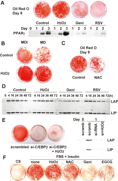
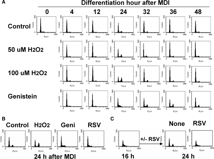
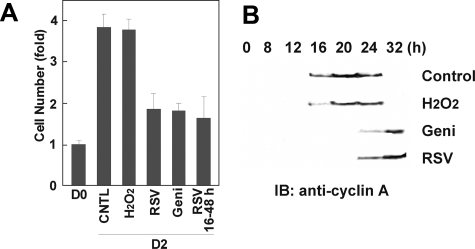
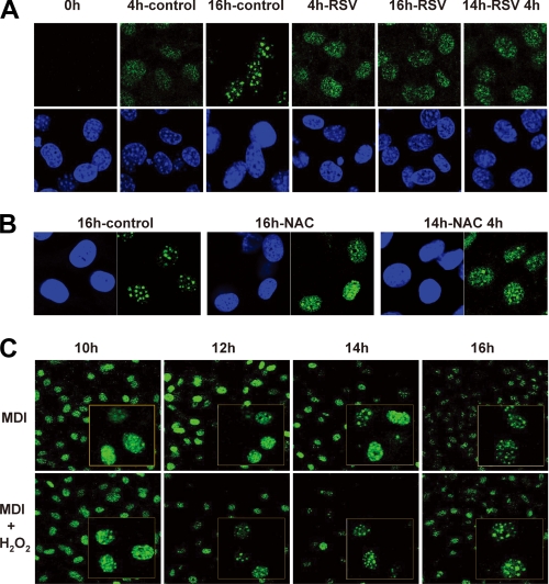
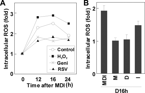
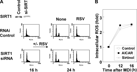
Growth-arrested 3T3-L1 preadipocytes rapidly express CCAAT/enhancer-binding protein-beta (C/EBPbeta) upon hormonal induction of differentiation. However, the DNA binding activity of C/EBPbeta is not activated until the cells synchronously reenter S phase during the mitotic clonal expansion (MCE) phase of differentiation. In this period, C/EBPbeta is sequentially phosphorylated by MAPK and glycogen synthase kinase-3beta, inducing C/EBPbeta DNA binding activity and transcription of its target genes. Because the DNA binding activity of C/EBPbeta is further enhanced by oxidation in vitro, we investigated how redox state affects C/EBPbeta DNA binding and MCE during adipogenesis. When 3T3-L1 cells were treated with H(2)O(2) and hormonal stimuli, differentiation was accelerated with increased expression of peroxisome proliferator-activated receptor gamma. Interestingly, cell cycle progression (S to G(2)/M phase) was markedly enhanced by H(2)O(2), whereas antioxidants caused an S phase arrest during the MCE. H(2)O(2) treatment resulted in the early appearance of a punctate pattern observed by immunofluorescent staining of C/EBPbeta, which is a hallmark for C/EBPbeta binding to regulatory elements, whereas a short antioxidant treatment rapidly dispersed the centromeric localization of C/EBPbeta. Consistently, reactive oxygen species production was increased during 3T3-L1 differentiation. Our results indicate that redox-induced C/EBPbeta DNA binding activity, along with the dual phosphorylation of C/EBPbeta, is required for the MCE and terminal differentiation of adipocytes.


Figures

FIGURE 1.
FIGURE 1.
The effects of ROS on differentiation of 3T3-L1 preadipocytes. Two-day postconfluent 3T3-L1 preadipocytes were induced to differentiate with the standard hormonal regimen (IBMX, dexamethasone, and insulin) in the presence or absence of H2O2 (100 μm), genistein (50 μm), resveratrol (50 μm) (A), or N-acetyl cysteine (10 mm) (B). After 2 days, all groups were fed with FBS, DMEM, and insulin for another 2 days and then with FBS + DMEM. On day 8, cells were fixed, and cytoplasmic triglyceride was stained with oil red O. In addition, whole cell extracts from day 1, 2, and 3 adipocytes were prepared and subjected to immunoblotting with antibody against PPARγ. C, cells were differentiated under the standard regimen (IBMX, dexamethasone, and insulin; MDI) or an incomplete regimen, lacking insulin (MD). D, cell lysates prepared from differentiating preadipocytes were subjected to SDS-PAGE and immunoblotted with antibody against C/EBPβ. LAP and LIP, 38-kDa liver-activating protein and 18-kDa liver-inhibiting protein, respectively. E, 3T3-L1 cells were treated with C/EBPβ siRNA at about 70% confluence using Lipofectamine RNAi/MAX reagent. After 24 h, medium was replaced with fresh DMEM containing 10% calf serum for 24 h before the induction of differentiation. Knockdown of C/EBPβ was verified by immunoblotting, and cells were differentiated with or without H2O2 (2-day treatment) and subjected to oil red O staining at day 8. F, 3T3-F442A cells were differentiated in the presence or absence of H2O2 (10 μm), genistein (50 μm), epigallocatechin 3-gallate (EGCG; 50 μm), or N-acetyl cysteine (10 mm).
FIGURE 2.
FIGURE 2.
FACS analysis during MCE of adipocyte differentiation. Cells were induced to differentiate in the presence or absence of H2O2, genistein (50 μm), or resveratrol (50 μm), and at the times indicated, cells were trypsinized and fixed. Changes in DNA content were analyzed by FACS using a FACSCalibur flow cytometry system, and data were analyzed using CellQuest software. FACS analysis was performed during 0–72 h of differentiation induction (A) and after 24 h of induction (B). C, cells were differentiated with the standard protocol for 16 h, after which resveratrol (50 μm) was added to the medium. FACS analysis was done at 24 h after induction of differentiation.
FIGURE 3.
FIGURE 3.
Effects of ROS on cell number and expression of cyclin A during MCE. A, on day 2 of differentiation, cells were trypsinized and subjected to cell counting. Cells were treated with H2O2 (100 μm), resveratrol (50 μm), or genistein (50 μm) for 48 or 32 h, as indicated. B, cell lysates were prepared at the times indicated and subjected to SDS-PAGE and immunoblotting (IB) to measure the expression of cyclin A.
FIGURE 4.
FIGURE 4.
Effects of ROS on intracellular localization of C/EBPβ during MCE. 3T3-L1 preadipocytes were induced to differentiate with the standard protocol in the presence or absence of H2O2 (100 μm), resveratrol (50 μm), or N-acetyl cysteine (10 mm), as indicated. Cells were fixed and subjected to immunofluorescence analysis with antibody against C/EBPβ and 4′,6-diamidino-2-phenylindole. Fluorescence images were obtained by confocal microscopy. Resveratrol (A) and N-acetyl cysteine (B) prevented the characteristic punctate pattern at 16 h of induction and also rapidly dispersed the centromeric localization after short term treatment (14–18 h of induction). C, H2O2 caused the early appearance of a punctate pattern in immunofluorescent stainings.
FIGURE 5.
FIGURE 5.
DNA-binding of C/EBPβ protein, in vivo and in vitro. A, chromatin immunoprecipitation (IP) analysis using the PPARγ promoter. At the indicated times after induction of differentiation with or without H2O2 (100 μm) or resveratrol (50 μm), cells were cross-linked with formaldehyde, the DNA was fragmented, and the chromatin-associated DNA was immunoprecipitated with IgG or antibodies against C/EBPβ. PCR amplification of the DNA fragments was conducted with specific primers corresponding to the PPARγ2 promoter sequence. Relative -fold changes of binding were calculated using a densitometer. B, EMSA of recombinant C/EBPβ protein treated by H2O2. Recombinant protein doubly phosphorylated by MAPK and GSK3β was treated with the indicated concentrations of H2O2 and/or 5 mm DTT and subjected to nonreducing SDS-PAGE, without DTT, and immunoblotting (IB) with anti-C/EBPβ antibody (top). The same reaction using 1 mm H2O2 was used for EMSA, except the reaction was terminated with 20 mm iodoacetamide at the indicated times, and 5 ng of protein was used for the binding reaction with the C/EBP site (bottom). DTT was added as marked (0–5 mm).
FIGURE 6.
FIGURE 6.
ROS production during MCE in adipogenesis. ROS were detected with the peroxide-sensitive fluorophore DCF-DA. At the times indicated, cells were incubated with DCF-DA (10 μm) for 1 h and analyzed by the FACSCalibur flow cytometry system; data are presented as relative intracellular ROS levels. A, ROS levels during MCE in the presence or absence of H2O2 (100 μm), genistein (50 μm), or resveratrol (50 μm); B, 3T3-L1 preadipocytes were treated with IBMX (M), dexamethasone (D), or insulin (I), respectively; ROS production was measured, as described above.
FIGURE 7.
FIGURE 7.
Resveratrol effects are independent of SIRT1 pathway. A, 3T3-L1 preadipocytes were subjected to RNA interference against siSIRT1. A ∼50% reduction of SIRT1 expression was confirmed by Western blotting, and FACS showed that SIRT1 knockdown did not affect the S phase accumulation of cells driven by resveratrol. B, ROS measurement during MCE from cells treated with aminoimidazole carboxamide ribonucleotide (AICAR) (1 mm) or sirtinol (20 μm).

References

    1. MacDougald, O. A., and Lane, M. D. (1995) Annu. Rev. Biochem. 64 345-373 - PubMed
    1. Farmer, S. R. (2006) Cell Metab. 4 263-273 - PMC - PubMed
    1. Rosen, E. D., Walkey, C. J., Puigserver, P., and Spiegelman, B. M. (2000) Genes Dev. 14 1293-1307 - PubMed
    1. Lane, M. D., Tang, Q. Q., and Jiang, M. S. (1999) Biochem. Biophys. Res. Commun. 266 677-683 - PubMed
    1. Tang, Q. Q., and Lane, M. D. (1999) Genes Dev. 13 2231-2241 - PMC - PubMed

Publication types

MeSH terms

LinkOut - more resources