. 2009 Feb 23;184(4):501-13.
doi: 10.1083/jcb.200808042.

Wld S requires Nmnat1 enzymatic activity and N16-VCP interactions to suppress Wallerian degeneration

Affiliations

Wld S requires Nmnat1 enzymatic activity and N16-VCP interactions to suppress Wallerian degeneration

Michelle A Avery et al. J Cell Biol. .

Abstract

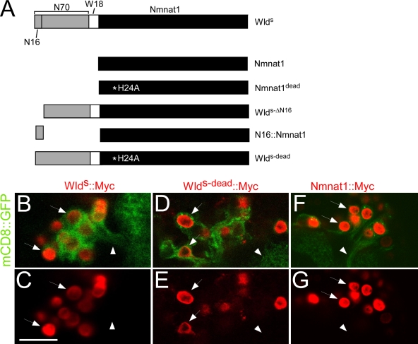
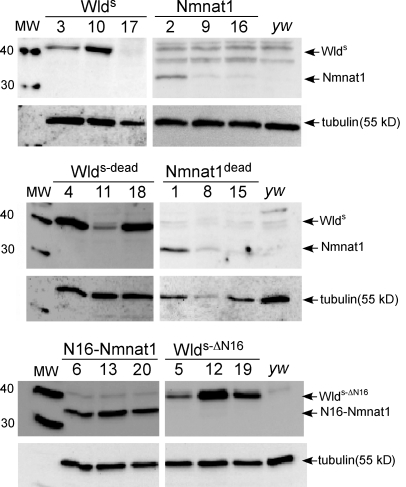
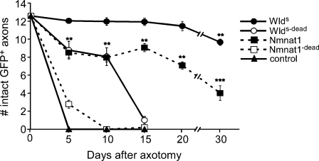
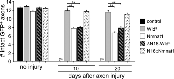
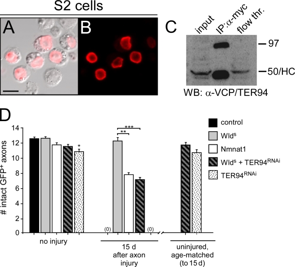
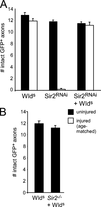
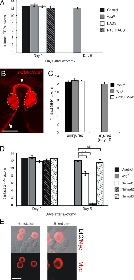
Slow Wallerian degeneration (Wld(S)) encodes a chimeric Ube4b/nicotinamide mononucleotide adenylyl transferase 1 (Nmnat1) fusion protein that potently suppresses Wallerian degeneration, but the mechanistic action of Wld(S) remains controversial. In this study, we characterize Wld(S)-mediated axon protection in vivo using Drosophila melanogaster. We show that Nmnat1 can protect severed axons from autodestruction but at levels significantly lower than Wld(S), and enzyme-dead versions of Nmnat1 and Wld(S) exhibit severely reduced axon-protective function. Interestingly, a 16-amino acid N-terminal domain of Wld(S) (termed N16) accounts for the differences in axon-sparing activity between Wld(S) and Nmnat1, and N16-dependent enhancement of Nmnat1-protective activity in Wld(S) requires the N16-binding protein valosin-containing protein (VCP)/TER94. Thus, Wld(S)-mediated suppression of Wallerian degeneration results from VCP-N16 interactions and Nmnat1 activity converging in vivo. Surprisingly, mouse Nmnat3, a mitochondrial Nmnat enzyme that localizes to the cytoplasm in Drosophila cells, protects severed axons at levels indistinguishable from Wld(S). Thus, nuclear Nmnat activity does not appear to be essential for Wld(S)-like axon protection.


Figures

Figure 1.
Figure 1.
Constructs used to dissect WldS functional domains and localization in Drosophila neurons. (A) All constructs were generated from mouse WldS, which contains N70, the first 16 amino acids (N16) that encode the VCP-binding site, W18, an 18–amino acid linker, and full-length Nmnat1. Nmnat1dead harbors an H24A mutation; WldSΔN16 lacks the N16 domain; N16-Nmnat1 is N16 tethered to the N terminus of Nmnat1; WldS-dead is full-length WldS with an H24A mutation in Nmnat1. See Materials and methods for details. (B–G) UAS-WldS–Myc (B and C), UAS-WldS-dead–Myc (D and E), and UAS-Nmnat1–myc (F and G) were expressed with UAS-mCD8–GFP in projection neurons using GH146-Gal4. The arrows point to nuclei, and the arrowheads point to axons. Bar, 12.14 µm.
Figure 2.
Figure 2.
WldS variant proteins are stably expressed in Drosophila neurons. Three independent UAS-driven transgene insertion lines for each WldS variant molecule were crossed to elav-Gal4, and protein extracts from heads were assayed for expression using α-Nmnat1 antibodies. Multiple lines for each construct were found to express detectable levels of protein. The expression of each of these molecules in ORNs with OR22a-Gal4 did not lead to expression of proteins at levels detectable by Western blot analysis (not depicted). Molecular masses are shown in kilodaltons. MW, molecular weight.
Figure 3.
Figure 3.
WldS requires N70 and Nmnat1 biosynthetic activity for maximal protection of severed axons in vivo. UAS-regulated versions of WldS variants were expressed in OR22a+ ORNs with OR22a-Gal4, axons were severed, and the number of remaining intact GFP+ axons was scored at the time points indicated. Two to four independent insertion lines were tested for each UAS-regulated WldS variant molecule (Fig. S1, available at http://www.jcb.org/cgi/content/full/jcb.200808042/DC1); n ≥ 10 for individual lines; data were subsequently pooled and are presented here. For Nmnat1, **, P < 0.01 and ***, P < 0.001 (Nmnat1 vs. WldS at corresponding time points). For WldS, **, P < 0.01 (day 0 vs. day 30). Error bars represent SEM.
Figure 4.
Figure 4.
N16 is the key domain in the N terminus of WldS that is essential for WldS-like axon protection. WldS, Nmnat1, ΔN16-WldS, and N16-Nmnat1 were expressed in ORNs using OR22a-Gal4, axons were severed, and the number of remaining intact GFP+ severed axons were scored at the time points indicated. n ≥ 10 for all genotypes; **, P < 0.01. Error bars represent SEM.
Figure 5.
Figure 5.
WldS localizes to the nucleus in Drosophila S2 cells and associates with TER94, and TER94 is essential for WldS-like levels of axon protection. (A and B) S2 cells were transfected with WldS-myc and stained with α-Myc antibodies, and α-Myc immunoreactivity localized to the nucleus. (C) Immunoprecipitation (IP) from WldS-Myc–transfected cell lysates with α-Myc antibodies led to the purification of a 97-kD isoform of Ter94. HC, heavy chain from α-Myc IgG; WB, Western blot. (D) UAS-Ter94RNAi was driven in OR22a+ ORNs in the presence or absence of WldS. n ≥ 10 for all genotypes; *, P < 0.05; **, P < 0.01; ***, P < 0.001. Error bars represent SEM. Bar, 13.56 µm.
Figure 6.
Figure 6.
Sir2 is not required for WldS-mediated protection of severed axons, and membrane-tethered WldS fails to suppress axon degeneration. (A) UAS-Sir2RNAi was driven in OR22a+ ORNs with OR22a-Gal4 in the presence or absence of WldS, and axons were assayed 15 d after injury. (B) The requirements for Sir2 in WldS-mediated axon protection were assayed 15 d after injury in WldS-expressing axons and WldS axons that lacked sir2 (sir24.5/sir25.26). WldS, n = 10; Sir2−/− + WldS, n = 6. Error bars represent SEM.
Figure 7.
Figure 7.
Axon-protective function of NAD+-producing enzymes is specific to Nmnat1 and -3, and Nmnat3 protects as well as WldS. (A) NADS and N16-NADS were expressed in ORNs using OR22a-Gal4, and axons were severed and analyzed for protection 5 d after axotomy. (B) Localization of mCD8-WldS was assayed using α-CD8 antibodies, and expression of mCD8-WldS was driven in ORNs using the OR22a-Gal4 driver. Axons are indicated projecting to glomeruli (arrowhead) and across the midline (arrow), and OR22a+ ORN-innervated glomeruli are shown with dashed lines. (C) Membrane-tethered WldS (mCD8-WldS) was assayed for axon-protective function 10 d after axotomy. n ≥ 10 for all genotypes. (D) Nmnat2 and -3 were driven by OR22a-Gal4 in ORNs to assay for protective function 5 d after axotomy. n ≥ 10 for all genotypes; **, P < 0.01; ***, P < 0.001. (E) S2 cells were transfected with Nmnat2-myc and Nmnat3-myc and stained with α-myc. In both cases, α-myc immunoreactivity was localized to the cytoplasm but not the nucleus. DIC, differential interference contrast. Error bars represent SEM. Bars: (B) 27.47 µm; (E) 14.71 µm.

References

    1. Araki T., Sasaki Y., Milbrandt J. 2004. Increased nuclear NAD biosynthesis and SIRT1 activation prevent axonal degeneration.Science. 305:1010–1013 - PubMed
    1. Beirowski B., Adalbert R., Wagner D., Grumme D.S., Addicks K., Ribchester R.R., Coleman M.P. 2005. The progressive nature of Wallerian degeneration in wild-type and slow Wallerian degeneration (WldS) nerves.BMC Neurosci. 6:6. - PMC - PubMed
    1. Bittner G.D. 1991. Long-term survival of anucleate axons and its implications for nerve regeneration.Trends Neurosci. 14:188–193 - PubMed
    1. Coleman M. 2005. Axon degeneration mechanisms: commonality amid diversity.Nat. Rev. Neurosci. 6:889–898 - PubMed
    1. Coleman M.P., Perry V.H. 2002. Axon pathology in neurological disease: a neglected therapeutic target.Trends Neurosci. 25:532–537 - PubMed

Publication types

Substances