. 2009 Mar 10;119(9):1241-52.
doi: 10.1161/CIRCULATIONAHA.108.783852. Epub 2009 Feb 23.

Long-term cardiac-targeted RNA interference for the treatment of heart failure restores cardiac function and reduces pathological hypertrophy

Affiliations

Long-term cardiac-targeted RNA interference for the treatment of heart failure restores cardiac function and reduces pathological hypertrophy

Lennart Suckau et al. Circulation. .

Abstract

Background: RNA interference (RNAi) has the potential to be a novel therapeutic strategy in diverse areas of medicine. Here, we report on targeted RNAi for the treatment of heart failure, an important disorder in humans that results from multiple causes. Successful treatment of heart failure is demonstrated in a rat model of transaortic banding by RNAi targeting of phospholamban, a key regulator of cardiac Ca(2+) homeostasis. Whereas gene therapy rests on recombinant protein expression as its basic principle, RNAi therapy uses regulatory RNAs to achieve its effect.

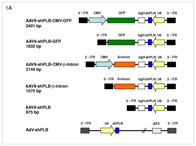
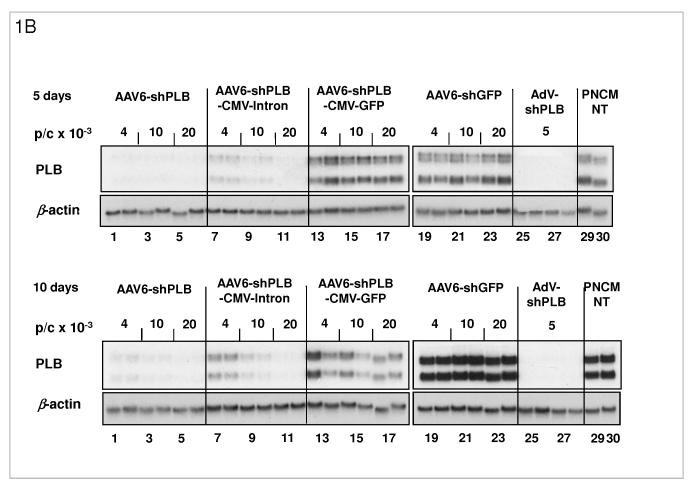
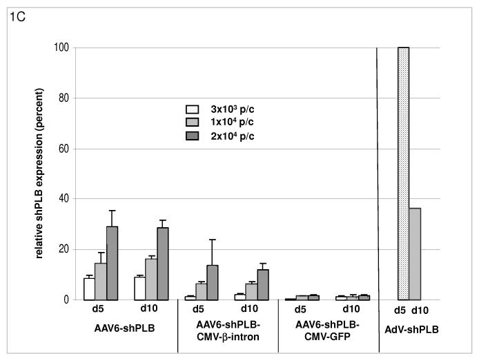
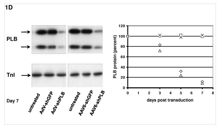
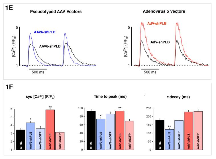
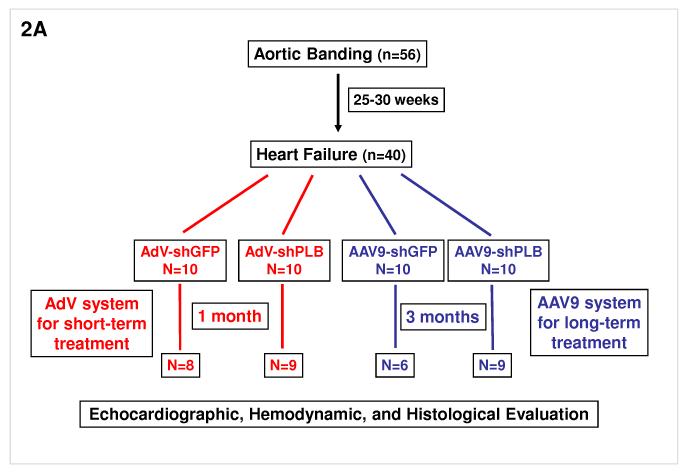
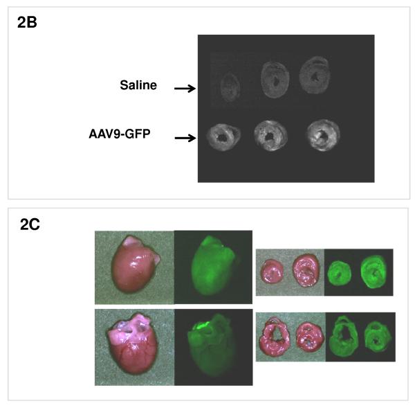
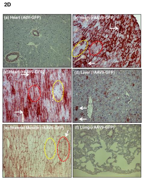
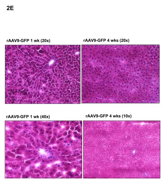
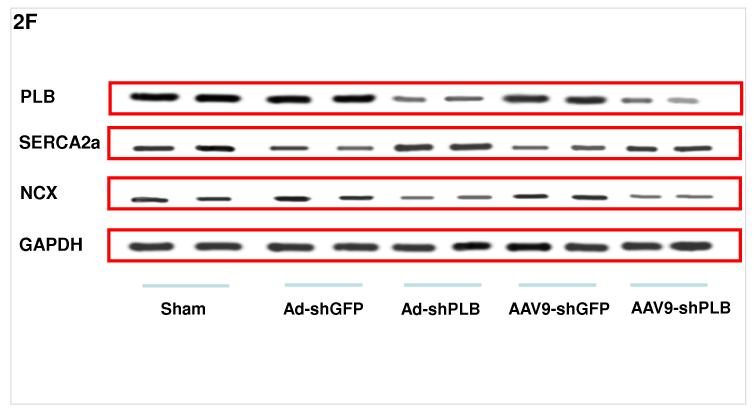
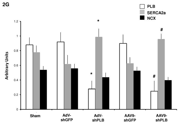
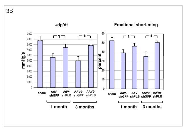
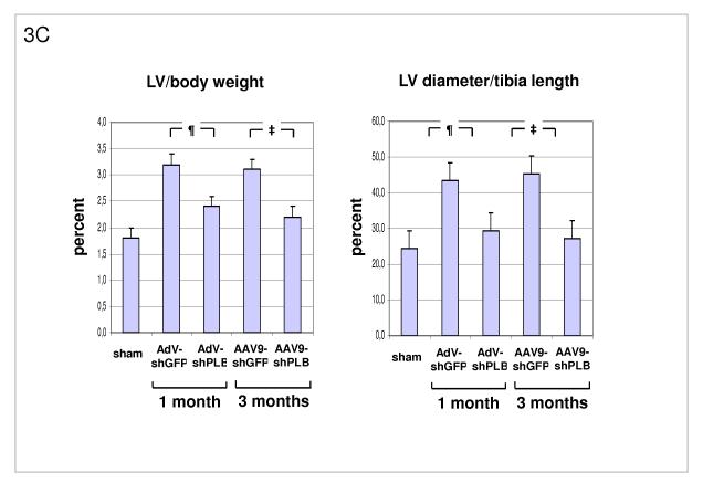
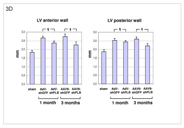
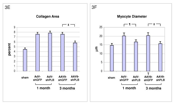
Methods and results: We describe structural requirements to obtain high RNAi activity from adenoviral and adeno-associated virus (AAV9) vectors and show that an adenoviral short hairpin RNA vector (AdV-shRNA) silenced phospholamban in cardiomyocytes (primary neonatal rat cardiomyocytes) and improved hemodynamics in heart-failure rats 1 month after aortic root injection. For simplified long-term therapy, we developed a dimeric cardiotropic adeno-associated virus vector (rAAV9-shPLB) to deliver RNAi activity to the heart via intravenous injection. Cardiac phospholamban protein was reduced to 25%, and suppression of sacroplasmic reticulum Ca(2+) ATPase in the HF groups was rescued. In contrast to traditional vectors, rAAV9 showed high affinity for myocardium but low affinity for liver and other organs. rAAV9-shPLB therapy restored diastolic (left ventricular end-diastolic pressure, dp/dt(min), and tau) and systolic (fractional shortening) functional parameters to normal ranges. The massive cardiac dilation was normalized, and cardiac hypertrophy, cardiomyocyte diameter, and cardiac fibrosis were reduced significantly. Importantly, no evidence was found of microRNA deregulation or hepatotoxicity during these RNAi therapies.

Conclusions: Our data show for the first time the high efficacy of an RNAi therapeutic strategy in a cardiac disease.


Figures

Fig. 1
Fig. 1. RNAi Vectors for HF Therapy
A: Maps of the RNAi vector genomes. Self-complementary “dimeric” AAV genomes (rAAV2) were pseudotyped into rAAV6 or rAAV9 capsids for studies in cardiomyocytes in vitro or HF rats in vivo, respectively. rAAV-shPLB has the same U6-shRNA transcription system as AdV-shPLB and the corresponding shGFP control vectors. Two further rAAV vectors carried CMV-GFP or CMV-β-intron expression cassettes in head-to-head orientation with the U6-shPLB sequence and separated from them by a bovine growth hormone (bgH) terminal signal. To assess the influence of the CMV promoter on vector function the CMV-free variants rAAV-shPLB-GFP and rAAV-shPLB-β-intron were constructed. Please note that the genomes in the rAAV6 vectors for in vitro work are identical to those shown for the rAAV9 vectors shown here. B: Comparison of the target silencing efficacy of shRNA vectors in NRCMs. Cells were harvested 5 (upper part) or 10 days (lower part), respectively, after treatment with the respective vector at the dose in particles per cell (p/c) given above the lanes. Northern blots were then carried out using a rat PLB-specific probe. To confirm equal RNA loading the blots were striped and rehybridized with a β-actin-specific probe. Lanes 1 to 18 show dose dependency of RNAi-mediated PLB-mRNA downregulation for the rAAV-based vectors rAAV-shPLB (lane 1-6), rAAV-shPLB-CMV- β-intron (lane 7-12), and rAAV-shPLB-CMV-GFP (lane 13-18). Lanes 19-24 show as a control for unspecific shRNA effects PLB-mRNA expression after treatment with rAAV-shGFP which generates an shRNA sequence targeting GFP (lanes 19-24). There was no difference towards untreated cells (lanes 29,30). For comparison with rAAV the adenovector AdV-shPLB (lanes 25-28) was used. PLB-mRNA was ≥98% ablated until day 10 by rAAV-shPLB at the lowest dose of 4×10³ p/c (lanes 1,2), similar to AdV-shPLB (lanes 25-28). Incorporation of a CMV-GFP expression cassette in the rAAV-shPLB vector (lanes 7-12) to provide this vector with a tag which is easily detectable by in vivo imaging, led to strong GFP expression in infected cells (not shown) but unexpectedly abolished its PLB gene silencing effect. Incorporation of a CMV-β-intron cassette (lanes 12-18) had a similar but less pronounced effect. We therefore used only rAAV9-shPLB vs rAAV9-shGFP and AdV-shPLB vs AdV-shGFP for in vivo therapy (Fig. 2a-g and 3a-f). C: The cellular shRNA levels produced by the vectors from panel B. In the presence of a CMV-GFP cassette shPLB production was abolished (lanes 13-18), whereas the U6-shPLB vector without additional sequences showed stable expression over 5 (lanes 1-3) and 10 days (lanes 4-6). AdV-shPLB generated very high shPLB levels on day 5 which then declined rather rapidly in NRCMs. Studies with further vectors (Suppl. Fig. 1a-c) showed that interaction of the CMV promotor with the shRNA-transcribing polymerase type III U6 promotor apparently disturbs shRNA transcription from the respective AAV vectors. D: On the left a Western blot analysis of PLB protein during treatment of NRCMs and on the right its quantitation on days 3, 5, and 7 after vector addition is shown. AdV-shPLB and rAAV6-shPLB resulted on day 7 in downregulation of cellular PLB to 9 % and 13 %, respectively, of baseline. E: [Ca2+]i transients in NRCMs during AAV9-shPLB treatment showed significantly higher amplitudes and accelerated transient kinetics (shortened TTP and τ) compared to the AAV9-shGFP group with transients indistinguishable from untreated cells. AdV-shPLB treatment also resulted in a significantly higher amplitude than in AdV-shGFP controls. In contrast to the AAV9 groups, TTP was prolonged in the AdV-shPLB vs. AdV-shGFP group which displayed no difference in τ. F: Statistical evaluation of the [Ca2+]i transients. * denotes p<0.05 and ** p<0.01. Additional studies of sarcoplasmic reticulum (SR) Ca2+ loading in the adenovirus groups (measured by rapid caffeine addition) were performed (Suppl. Fig. 1d,e) and showed increased SR Ca2+ loading and fractional Ca2+ release from the SR in AdV-shPLB vs. AdV-shGFP group.
Fig. 2
Fig. 2. Protocol for RNAi Therapy of HF
A: Animals for the in vivo RNAi therapy study were divided in two groups: one of 56 animals with aortic banding (TAB) and a second of 12 sham-operated. In the TAB animals we waited 25-30 weeks for them to develop LV dilatation and a decrease in fractional shortening by 25% (echocardiography) prior to cardiac RNAi vector transfer. From 56 TAB animals 40 survived and were further divided to receive AdV-shGFP (n=10), AdV-shPLB (n=10), rAAV9-shGFP (n=10), or rAAV9-shPLB (n=10). 3×10¹⁰ pfu of each AdV were injected in 200 μl of solution. For experiments with rAAV9, tail vein injection was done using 5×10¹¹ genomes of either vector. Outcome evaluation by echocardiography, tip catheter, morphometry, and histology was after 1 month in the AdV and after 3 months in the rAAV9 groups (Fig. 3a-f). In the AdV groups 8/10 and 9/10 animals survived after 1 month. After 3 months 9/10 survived in the rAAV-shPLB and 6/10 in the rAAV-shGFP group. B: Rats were injected i.v. with an rAAV9-GFP vector expressing green florescent protein, or with saline. 1 month later the hearts were removed and visualized by GFP imaging which showed a grossly homogeneous signal in cardiac cross sections in the (lower) rAAV9-GFP group, and no signal in the (upper) saline group. C: An overview on GFP fluorescence from hearts from rAAV9-GFP-treated rats reaching 90% of surface area at 1 month. D: Immunhistochemical staining of GFP in different organs 1 month after i.v. injection of rAAV9-GFP. Whereas after i.v. injection of an adenoviral vector (AdV-GFP) no GFP was detected in the heart (a), rAAV9-GFP treatment resulted in strong GFP expression (b)(c) which was grossly homogeneous. Few areas are completely devoid of GFP immunoreactivity (encircled yellow), others show homogeneous cytoplasmic staining (encircled red). Staining is particularly dense at sites where high expression over 1 month has obviously resulted in the formation of precipitates (white arrows) of GFP which is stable in cells, in contrast to shRNA generated from RNAi vectors. An average of 70% of cardiomyocytes were positive by immunohistochemistry, with variability of expression among individual cells. (e) shows skeletal muscle with faint staining of a fraction of cells, whereas the liver shows prominent signal of individual cells only (d). No signal was visible in the lungs. Further data on AAV9 distribution are given in Suppl. Fig. 2c with GFP quantitation by Western blot analyses, documenting highest affinity of rAAV9-GFP expression for the heart. Liver and skeletal muscle showed low and the lungs only very faint expression. Suppl. Fig. 2ab document specificity of the GFP staining. E: Haematoxylin-eosin staining of livers 1 week and 4 weeks after i.v. injection of rAAV9-GFP shows no evidence of toxicity. rAAV-shRNA vector also resulted in no hepatotoxicity. F: Representative Western blots showing a significant decrease of cardiac PLB protein after 1 month of AdV-shPLB and 3 months of rAAV9-shPLB therapy compared to the shGFP control groups. The NCX and GAPDH protein remained unchanged. SERCA2a was decreased in the shGFP groups which were in heart failure as compared to sham, whereas SERCA2a was significantly increased in both shPLB groups. G: Statistical evaluation of Western blots from the different treatment groups. * denotes p<0.05 compared to AdV-shGFP, # p<0.05 compared to rAAV9-shGFP.
Fig. 3
Fig. 3. Functional and Morphological Effects of RNAi Therapy
A: Influence of RNAi treatments on parameters of diastolic LV function. The high LV filling pressure (LVEDP) in rats after TAB was significantly lowered by shPLB vectors (lanes 3,5) compared to shGFP controls (lanes 2,4). The maximal rate of pressure fall (−dP/dt) was significantly increased by shPLB treatment, also the isovolumetric relaxation time constant Tau (Suppl. Fig. 3a). Values were restored to normal range (lane 1) after 3 months of rAAV-shPLB therapy (lane 5). ¶ denotes p<0.05 for AdV-shPLB vs. AdV-shGFP, ‡ p<0.05 for rAAV9-shPLB vs. rAAV9-shGFP. B: RNAi treatment effects on systolic function. Echocardiography showed normalized fraction shortening (FS) after 3 months of rAAV-shPLB therapy, whereas FS improvement was also significant but less pronounced for AdV-shPLB. The maximal rate of pressure rise (+dP/dt) was improved compared to controls, also the systolic pressure (LVSP)(Suppl. Fig. 3b). C: Morphometry post mortem showing marked LV hypertrophy induced by TAB (lanes 2,4) with LV weight (Suppl. Fig. 3c) and LV/body weight (LV/BW) ratio ≈ 2/3 thirds above baseline (lane 1). There was also marked LV dilation (LV diameter/tibia length ratio). The latter was reduced to within the normal range after 1 or 3 months of therapy with AdV-shPLB or rAAV9-shPLB (lanes 3,5). Cardiac hypertrophy was significantly reduced by both vector types. D: A summary of echocardiographic data on cardiac morphology which corroborate the morphometric findings in panel C (see also Suppl. Fig. 3d). E: Cardiac collagen content in HF animals after RNAi therapy. After therapy with AdV-shPLB there was no decrease in fibrosis at 1 month, whereas rAAV9-shRNA treatment resulted in significantly reduced fibrosis at 3 months. The control vectors had no effect. F: Both treatment modes induced a significant decrease in cardiomyocyte diameters.

References

    1. Schmidt U, Hajjar RJ, Kim CS, Lebeche D, Doye A, Gwathmey J. Human heart failure: cAMP stimulation of SR Ca2+-ATPase activity and phosphorylation level of phospholamban. Am J Physiol. 1999;277:H474–480. - PubMed
    1. MacLennan D, Kranias E. Phospholamban: A crucial regulator of cardiac contractility. Nature Reviews Molecular Cell Biology. 2003;4:566–576. - PubMed
    1. Hoshijima M, Ikeda Y, Iwanaga Y, Minamisawa S, Date M, Gu Y, Iwatate M, Li M, Wang L, Wilson J, Wang Y, Ross J, Chien K. Chronic suppression of heart-failure progression by a pseudophosphorylated mutant of phospholamban via in vivo cardiac rAAV gene delivery. Nature Medicine. 2002;8:864–871. - PubMed
    1. Iwanaga Y, Hoshijima M, Gu Y, Iwatate M, Dieterle T, Ikeda Y, Date MO, Chrast J, Matsuzaki M, Peterson KL, Chien KR, Ross J., Jr. Chronic phospholamban inhibition prevents progressive cardiac dysfunction and pathological remodeling after infarction in rats. J Clin Invest. 2004;113:727–736. - PMC - PubMed
    1. Eizema K, Fechner H, Bezstarosti K, Schneider-Rasp S, van der Laarse A, Wang H, Schultheiss H-P, Poller W, Lamers J. Adenovirus-based phospholamban-antisense-mRNA expression as a novel approach to improve cardiac contractile dysfunction - Comparison of a constitutive viral versus an endothelin-1-responsive cardiac promoter. Circulation. 2000;101:2193–2199. - PubMed

Publication types

MeSH terms