. 2009 May;182(1):41-54.
doi: 10.1534/genetics.109.101683. Epub 2009 Feb 23.

Alternative induction of meiotic recombination from single-base lesions of DNA deaminases

Affiliations

Alternative induction of meiotic recombination from single-base lesions of DNA deaminases

Siim Pauklin et al. Genetics. 2009 May.

Abstract

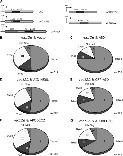
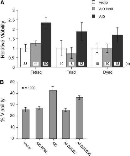
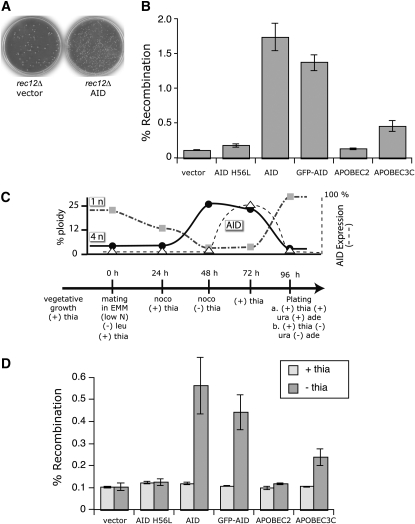
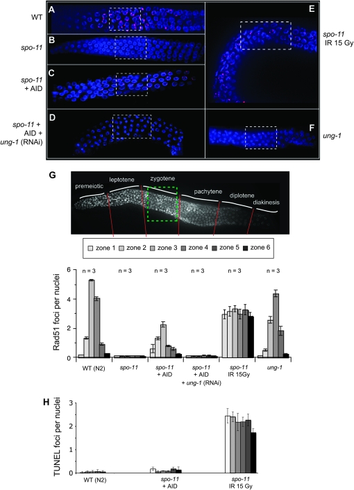
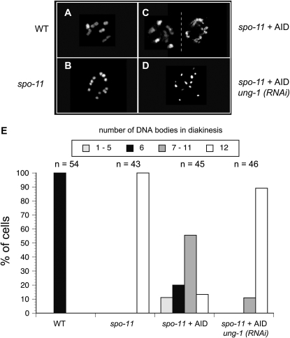
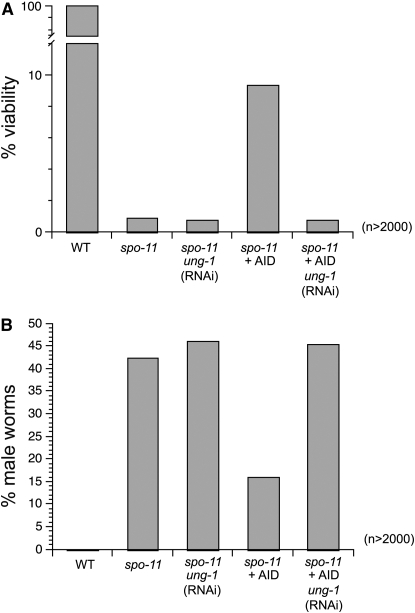
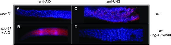
Meiotic recombination enhances genetic diversity as well as ensures proper segregation of homologous chromosomes, requiring Spo11-initiated double-strand breaks (DSBs). DNA deaminases act on regions of single-stranded DNA and deaminate cytosine to uracil (dU). In the immunoglobulin locus, this lesion will initiate point mutations, gene conversion, and DNA recombination. To begin to delineate the effect of induced base lesions on meiosis, we analyzed the effect of expressing DNA deaminases (activation-induced deaminase, AID, and APOBEC3C) in germ cells. We show that meiotic dU:dG lesions can partially rescue a spo11Delta phenotype in yeast and worm. In rec12 Schizosaccharomyces pombe, AID expression increased proper chromosome segregation, thereby enhancing spore viability, and induced low-frequency meiotic crossovers. Expression of AID in the germ cells of Caenorhabditis elegans spo-11 induced meiotic RAD-51 foci formation and chromosomal bivalency and segregation, as well as an increase in viability. RNAi experiments showed that this rescue was dependent on uracil DNA-glycosylase (Ung). Furthermore, unlike ionizing radiation-induced spo-11 rescue, AID expression did not induce large numbers of DSBs during the rescue. This suggests that the products of DNA deamination and base excision repair, such as uracil, an abasic site, or a single-stranded nick, are sufficient to initiate and alter meiotic recombination in uni- and multicellular organisms.


Figures

F<sc>igure</sc> 1.—
Figure 1.—
DNA deaminases partially restore tetrad formation in rec12Δ S. pombe. (A) Schematic representation of the constructs used in this study. Proteins were expressed under the control of thiamine-repressible nmt1 or nmt41 promoter. Expression is driven in the absence of thiamine. Amino acids essential for the formation of the catalytic domain (blue) are indicated as well as the substitution of His56 with Leu56 in human AID (AID), which renders the protein catalytically inactive. (B–G) Relative distribution of tetrads (I), triads (II), dyads (III), and missegregations (IV) that are formed during meiosis in a rec12Δ background transfected with an empty vector (B), with human AID (C), with a catalytically mutant human AID (D), with GFP-human AID (E), with human APOBEC2 (F), and with human APOBEC3C (G). Cells were transfected, grown in the presence of thiamine for 24 hr, washed, grown in the absence of thiamine for 6 hr, and crossed for 2 days on thiamine (−) plates. Tetrads, triads, dyads, and missegregations were quantitated by microscopy.
F<sc>igure</sc> 2.—
Figure 2.—
Deaminases increase spore viability after meiosis. (A) Ascus dissection analysis: relative viabilities of rec12Δ single spores from tetrads, triads, and dyads in control-transfected cells (white), in AID catalytic mutant expressing cells (orange), and in catalytically active AID expressing cells (red). The numbers of the spores that were isolated are indicated at the bottom of each column, and the viability of the vector-only rec12Δ strain was set to one. (B) Percentage of viability of random spore analysis, from rec12Δ cells expressing different deaminases during meiosis.
F<sc>igure</sc> 3.—
Figure 3.—
Meiotic recombination initiated by AID. (A) Representative plates showing the increase in recombinants after meiosis of rec12Δ cells transfected with empty vector (left plate) or with AID (right plate). Cells auxotrophic for either adenine (ade6) or uracil (ura4) were crossed on EMM low N and spores were plated on EMM media supplemented with phloxin B and in the absence or the presence of both adenine and uracil, to assess the relative number of recombinants compared to the total number of viable spores. Shown are the EMM plates lacking adenine and uracil. (B) Fold increase in ade6 ura4 recombinants after the expression of AID catalytic mutant, functional AID, GFP-AID, APOBEC2, or APOBEC3C during meiosis compared to the empty vector (set to one) control. (C) Time line of postreplicative induction of AID-induced recombination. Time points represent the relative timing of changing the media or the addition of the indicated compounds nocodazole (noco), leucine (leu), and thiamine (thia); + and − indicate the presence or the absence of the compounds starting from the corresponding time point. Cell ploidy, visualized with propidium iodide in FACS, is shown as a solid line (solid circles, 4n) and a shaded dashed-dotted line (shaded squares, 1n), and AID expression, visualized by GFP in FACS, is shown as a dashed line (based on Figure S2, open triangle). (D) DNA deaminase-mediated recombination events can be initiated after DNA replication. Cells were treated as indicated in the time line (C). The graph shows the recombinants obtained when AID, mutant (H56L) AID, and the APOBEC proteins were expressed after removal of thiamine (− thia) at a 48-hr time point compared to the results when deaminase expression is repressed during meiosis by thiamine (+ thia). For details see materials and methods.
F<sc>igure</sc> 4.—
Figure 4.—
AID induces RAD-51 foci during meiosis in the spo-11 worm. Representative pictures of germlines visualized for RAD-51 foci, using anti-RAD-51 antibodies (red) as well as DAPI staining to visualize the nuclei, are shown: (A) wild-type animals, (B) spo-11, (C) spo-11 worm expressing AID, (D) spo-11 worm expressing AID with RNAi treatment for ung-1, (E) spo-11 worm irradiated with 15 Gy and analyzed after 4 hr, and (F) ung-1 defective nematode. Dashed squares represent the proposed location of zygotene stage in the germline. The germlines were divided into six zones according to the developmental stage of the germline. (G) The number of RAD-51 foci per nucleus was counted in worm germlines with respective genotypes (A–F) in the six developmental zones (30 nuclei per zone per worm, analyzing three worms per genotype). (H) The number of TUNEL stains per nucleus in the six developmental zones was counted from the germline of the indicated worms (for details see Figure S3). The fold difference between spo-11 (IR 15 Gy) and spo-11 + AID was >20-fold and >50-fold for the wt.
F<sc>igure</sc> 5.—
Figure 5.—
AID alters the chromosome morphology during the diakinesis stage. Representative pictures are shown of DAPI-stained chromosomes during diakinesis from (A) wild type, (B) spo-11, (C) two nuclei of an AID-expressing spo-11 worm, and (D) an AID-expressing spo-11 worm after treatment with ung-1 (RNAi). (E) The numbers of DAPI-staining bodies per nucleus were counted for each of the corresponding genotypes from the indicated number of cells. Different shadings for the columns represent the numbers of DAPI-stained bodies as indicated on the top of the graph.
F<sc>igure</sc> 6.—
Figure 6.—
AID increases viability and hermaphrodite frequency in spo-11 worms. (A) Worm viabilities for the indicated genotypes were assessed by counting viable offspring and dead eggs in a brood from young adults. (B) The frequencies of males among viable progeny from the sample in A were assessed by microscopic analysis of tail morphology and movement. A total of at least 2000 worms were counted per genotype (for extended data see Table 1).
F<sc>igure</sc> 7.—
Figure 7.—
Expression of AID and Ung during oogenesis. AID was detected with anti-AID antibodies (red) in (A) a spo-11 worm and (B) an AID-expressing spo-11 worm. UNG was detected with anti-Ung antibody (red) in (C) a wild-type worm germline and (D) a wild-type worm treated with ung-1 (RNAi). Nuclei were counterstained with DAPI.

Comment in

References

    1. Aoufouchi, S., A. Faili, C. Zober, O. D'Orlando, S. Weller et al., 2008. Proteasomal degradation restricts the nuclear lifespan of AID. J. Exp. Med. 205 1357–1368. - PMC - PubMed
    1. Arakawa, H., G. L. Moldovan, H. Saribasak, N. N. Saribasak, S. Jentsch et al., 2006. A role for PCNA ubiquitination in immunoglobulin hypermutation. PLoS Biol. 4 e366. - PMC - PubMed
    1. Bahler, J., T. Wyler, J. Loidl and J. Kohli, 1993. Unusual nuclear structures in meiotic prophase of fission yeast: a cytological analysis. J. Cell Biol. 121 241–256. - PMC - PubMed
    1. Barreto, V., B. Reina-San-Martin, A. R. Ramiro, K. M. McBride and M. C. Nussenzweig, 2003. C-terminal deletion of AID uncouples class switch recombination from somatic hypermutation and gene conversion. Mol. Cell 12 501–508. - PubMed
    1. Boulton, S. J., A. Gartner, J. Reboul, P. Vaglio, N. Dyson et al., 2002. Combined functional genomic maps of the C. elegans DNA damage response. Science 295 127–131. - PubMed

Publication types

MeSH terms

Substances