. 2009 May;182(1):121-32.
doi: 10.1534/genetics.109.100693. Epub 2009 Feb 23.

Sex-lethal facilitates the transition from germline stem cell to committed daughter cell in the Drosophila ovary

Affiliations

Sex-lethal facilitates the transition from germline stem cell to committed daughter cell in the Drosophila ovary

Johnnie Chau et al. Genetics. 2009 May.

Abstract

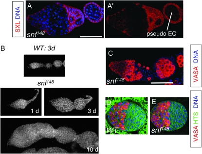
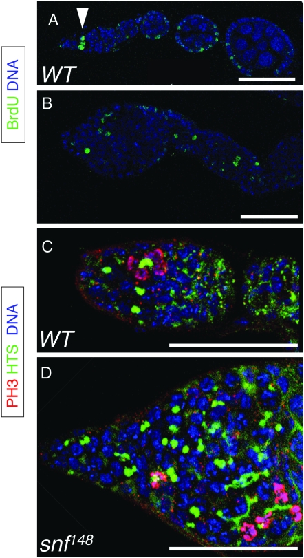
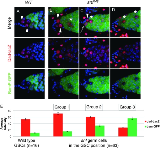
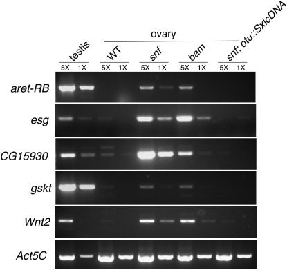
In Drosophila, the female-specific SEX-LETHAL (SXL) protein is required for oogenesis, but how Sxl interfaces with the genetic circuitry controlling oogenesis remains unknown. Here we use an allele of sans fille (snf) that specifically eliminates SXL protein in germ cells to carry out a detailed genetic and cell biological analysis of the resulting ovarian tumor phenotype. We find that tumor growth requires both Cyclin B and zero population growth, demonstrating that these mutant cells retain at least some of the essential growth-control mechanisms used by wild-type germ cells. Using a series of molecular markers, we establish that while the tumor often contains at least one apparently bona fide germline stem cell, the majority of cells exhibit an intermediate fate between a stem cell and its daughter cell fated to differentiate. In addition, snf tumors misexpress a select group of testis-enriched markers, which, remarkably, are also misexpressed in ovarian tumors that arise from the loss of bag of marbles (bam). Results of genetic epistasis experiments further reveal that bam's differentiation-promoting function depends on Sxl. Together these data demonstrate a novel role for Sxl in the lineage progression from stem cell to committed daughter cell and suggest a model in which Sxl partners with bam to facilitate this transition.


Figures

F<sc>igure</sc> 1.—
Figure 1.—
Sxl and bamP-GFP coexpression in the germarium. (A) Schematic of a wild-type germarium. The cell destined to become the oocyte is blue, and all other germline-derived cells are pink. Spectrosomes and fusomes are red. Cells of somatic origin are in shades of green and gray. The following abbreviations are used: GSC, germline stem cell; CB, cystoblast; fu, fusome; sp, spectrosome. (B, B′, and B″) Wild-type germarial tip from a female carrying the bamP-GFP reporter construct stained for GFP (green), SXL (red), and DNA (blue). Cytoplasmic SXL staining is prominent in two cell types: GSC cells, which do not express bamP-GFP, and CB cells, where bamP-GFP is expressed. As differentiation proceeds, bamP-GFP staining increases and cytoplasmic SXL staining decreases.
F<sc>igure</sc> 2.—
Figure 2.—
snf148 germline tumors. (A and A′) snf148/snf148 mutant germarium and adjoining pseudo-egg chamber (EC) stained for SXL (red) and DNA (blue). Bar, 50 μm. (B) DAPI-stained ovariole from a 3-day-old wild-type (WT) female (top) and a 1-day-old, a 3-day-old, and a 10-day-old snf148/snf148 mutant female (bottom). (C) snf148/snf148 mutant germarium and adjoining pseudo-egg chamber (EC) stained for VASA (red) and DNA (blue). The image is the same magnification as in A. (D and E) Control (D) and snf148/snf148 (E) gonads from third instar female larvae stained for VASA (red), HTS (green), and DNA (blue). Note that HTS staining labels both spectrosomes and the somatic cell membranes. In control gonads (D) the fat body surrounding the gonad is also green because of expression of a GFP marker, as described in materials and methods. The images in D and E are the same magnification.
F<sc>igure</sc> 3.—
Figure 3.—
Proliferating snf148 tumor cells are located throughout the germarium and in pseudo-egg chambers. (A and B) Ovarioles from wild-type (A) and snf148/snf148 females (B) stained for BrdU incorporation (green) to label cells in S-phase and for DNA (blue). In wild type, BrdU positive germ cells are limited to the tip of the germarium (arrowhead). Other cells that are observed to be BrdU positive include the somatic follicle cells that surround the egg chambers. In snf148 mutants, BrdU-positive germ cells are dispersed throughout the germarium and in the pseudo-egg chamber. Bars, 50 μm. (C and D) Germaria from wild-type (C) and snf148/snf148 females (D) stained for HTS (green), DNA (blue), and PH3 (magenta when merged with blue DNA stain). In wild type, interconnected germ cells in mitosis are judged to be in synchrony on the basis of the morphology of the PH3 staining chromosomes. In snf148 mutants, both single cells and groups of interconnected cells are found to be in mitosis. Bars, 50 μm.
F<sc>igure</sc> 4.—
Figure 4.—
CycB is required for snf148 tumor growth. (A and B) Germaria from wild-type (A) and snf148/snf148 mutant (B) females carrying the protein trap allele, P{PTT-GC}CycBCC01846 stained for GFP to detect the CYCB-GFP fusion protein (green) and DNA (blue). In wild type, CYCB-GFP expression is highest at the anterior of the germarium, where the dividing germ cells reside. Staining is also visible in the dividing somatic cells, including the follicle cells that surround the egg chamber. In snf148 mutant ovaries, CYCB-GFP expression is observed throughout the tumor. The images in A and B are the same magnification. (C and D) Ovaries from double-mutant snf148/snf148; CycB2/Df(2R)59AB females (C) or single-mutant snf148/+; CycB2/Df(2R)59AB females (D) stained for VASA (red) and DNA (blue) to show that the removal of CycB suppresses the snf tumor phenotype. While the majority of both double-mutant and control ovaries were found to be devoid of VASA staining cells, the images in C and D are examples of the few ovarioles that contained germ cells. The images in C and D are the same magnification.
F<sc>igure</sc> 5.—
Figure 5.—
GSC- and CB-specific markers are expressed throughout snf148 mutant ovaries. (A and B) Germaria from wild-type (A) and snf148/snf148 females (B) carrying the nuclear myc-Piwi reporter construct stained for MYC (red or magenta when merged with the blue DNA stain) and DNA (blue). In wild-type germ cells, PIWI is limited to two to three cells at the tip of the germarium. In snf tumors, PIWI-expressing cells are located throughout the tumor. Bar in A, 50 μm. The images in A–F are the same magnification. (C and D) Germaria from wild-type (C) and snf148/snf148 females (D) stained for PUM protein expression (green) and DNA (blue). In wild type, the cytoplasmic PUM protein is highly expressed in two to three cells at the tip of the germarium. In snf148 mutant germaria, PUM-expressing cells are located throughout the tumor. (E and F) Germaria from wild-type (A) and snf148/snf148 females (B) carrying the bamP-GFP reporter construct stained for GFP (green) and DNA (blue). In wild type, only CB cells and dividing cysts express bamP-GFP. Germ cells within the snf148 mutant germaria, however, continue to express bamP-GFP, even in the pseudo-egg chamber. Furthermore, in many cases we observed stained cells located at the tip of the germaria (see text and Figure 6). (G) Western blot of extracts made from ovaries dissected from wild-type and snf148/snf148 mutant females probed with a polyclonal antibody against the Bam-C protein and the U1-70K protein as a loading control.
F<sc>igure</sc> 6.—
Figure 6.—
TGF-β signaling reception in snf148 mutant germ cells. (A–D) Germaria from wild-type (A) and snf148/snf148 (B–D) females carrying both the dad-lacZ and bamP-GFP reporter constructs stained for β-galactosidase (red), GFP (green), and DNA (blue). Arrows and Arrowheads indicate the presumptive GSCs, identified by their position within the germarium and expression of dad-lacZ. In snf mutant germaria the additional β-galactosidase staining cells located elsewhere in the tumor are marked with an asterisk. (E) Quantification of β-galactosidase and GFP staining intensity in cells adjacent to the somatic cap cells. The average intensity (± SEM) is presented. Wild-type cells were all very similar to each other, with levels of β-galactosidase staining ranging from 40 to 80 and basal levels of GFP. snf148/snf148 mutant cells on the other hand varied and were divided into three groups on the basis of their staining patterns. Groups 1 and 2 all had levels of β-galactosidase staining that fell within the wild-type range, i.e., >40. This group was further subdivided into those that expressed GFP within the wild-type range, i.e., <20 (group 1), and those that had higher levels of GFP staining (group 2). Those cells with levels of β-galactosidase staining of <40 were placed in group 3.
F<sc>igure</sc> 7.—
Figure 7.—
snf148; bamΔ86 double-mutant analysis. (A–D) Germaria from wild-type (A), snf148/snf148; bamΔ86/+ (B), snf148/+; bamΔ86/bamΔ86 (C), and double-mutant snf148/snf148; bamΔ86/bamΔ86 (D) animals stained for HTS (green) and DNA (blue). In wild type (A) HTS labels the round spectrosomes (arrowhead, sp) and fusomes (arrowhead, fu). Note that HTS also labels somatic cell membranes. In snf mutants (B), round (arrowhead, R), short barbell-shaped (arrowhead, S), and longer branched structures (arrowhead, L) are visible throughout the germarium. In bam mutants (C), the majority of HTS staining material resembles round spectrosomes. In double mutants (D), the morphology of the spectrosomes/fusomes is more similar to the snf mutant phenotype with accumulation of short and elongated fusome-like structures. Bars, 50 μm. (E) Quantification of the spectrosome/fusome morphology observed in double-mutant and control germaria. For each genotype, the number and shape of the spectrosome/fusome-like structures in each germarium were recorded (sample size, 10 germaria per genotype). The structures were classified as (1) round (spherical and not protruding into other cells), (2) short (branches into one adjacent cell), or (3) long (branches into two or more adjacent cells).
F<sc>igure</sc> 8.—
Figure 8.—
Testis-enriched markers are expressed in both snf and bam mutant ovaries. RT–PCR analysis is shown of aret-RB, esg, CG15930, gskt, Wnt2, and Act5C mRNA levels in testis from wild-type animals and in ovaries from wild-type, snf148/snf148, bamΔ86/bamΔ86, and snf148/snf148; P{otu∷SxlcDNA} females. Act5C is used here as a loading control. To ensure that the mRNA measurements fall within a linear range of amplification, each RT reaction was serially diluted fivefold (1× and 5×). Similar results were obtained in three independent biological replicates.
F<sc>igure</sc> 9.—
Figure 9.—
A model for ovarian tumor formation. (A) In wild type the GSC cell (pink) divides asymmetrically to produce one daughter cell that will remain a stem cell and a second daughter cell destined to differentiate (pink and green striped) called a pre-CB cell because these cells express both GSC markers and BAM. Before the next division, the cell matures and GSC marker expression is attenuated (green cell). Here we propose that the SXL protein, which is highly expressed in the cytoplasm of both GSC and CB cells, partners with the newly expressed BAM protein, to antagonize the GSC-specific gene expression program. The mature CB cell then undergoes four rounds of mitosis to yield a 16-cell differentiated cyst. (B) In the absence of SXL or BAM protein the pre-CB cells continue to express GSC markers and misexpress a set of testis-specific markers (blue stripes). As a result, these differentiation-defective cells fail to mature and continue to divide symmetrically, forming a germline tumor.

References

    1. Bopp, D., L. R. Bell, T. W. Cline and P. Schedl, 1991. Developmental distribution of female-specific SEX-LETHAL proteins in Drosophila melanogaster. Genes Dev. 5 403–415. - PubMed
    1. Bopp, D., J. I. Horabin, R. A. Lersch, T. W. Cline and P. Schedl, 1993. Expression of the Sex-lethal gene is controlled at multiple levels during Drosophila oogenesis. Development 118 797–812. - PubMed
    1. Buszczak, M., S. Paterno, D. Lighthouse, J. Bachman, J. Planck et al., 2007. The Carnegie protein trap library: a versatile tool for Drosophila developmental studies. Genetics 175 1505–1531. - PMC - PubMed
    1. Casanueva, M. O., and E. L. Ferguson, 2004. Germline stem cell number in the Drosophila ovary is regulated by redundant mechanisms that control DPP signaling. Development 131 1881–1890. - PubMed
    1. Casper, A., and M. Van Doren, 2006. The control of sexual identity in the Drosophila germline. Development 133 2783–2791. - PubMed

Publication types

MeSH terms