. 2009 Feb-Mar;32(2):145-56.
doi: 10.1097/CJI.0b013e318193d31e.

Targeting the intratumoral dendritic cells by the oncolytic adenoviral vaccine expressing RANTES elicits potent antitumor immunity

Affiliations

Targeting the intratumoral dendritic cells by the oncolytic adenoviral vaccine expressing RANTES elicits potent antitumor immunity

Natalia Lapteva et al. J Immunother. 2009 Feb-Mar.

Abstract

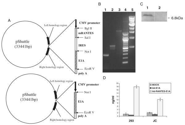
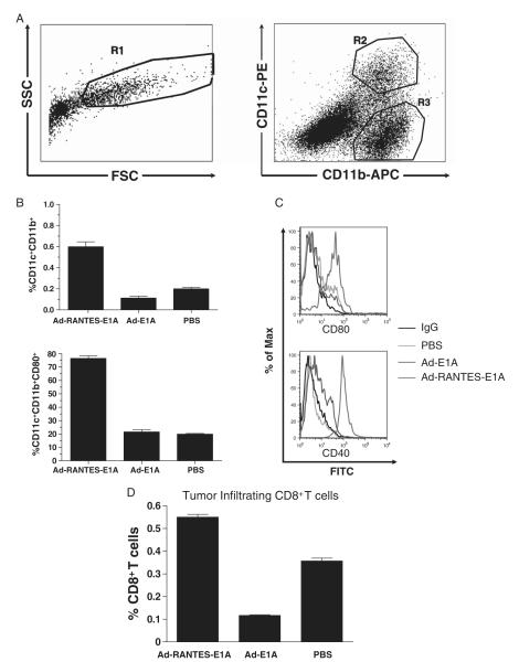
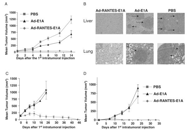
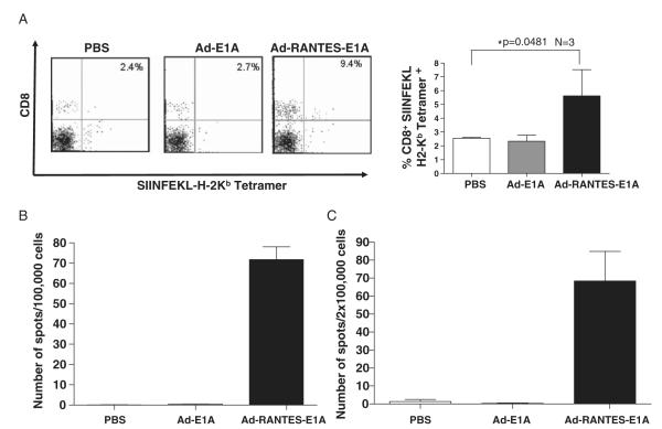
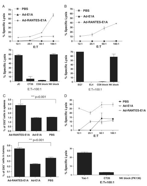
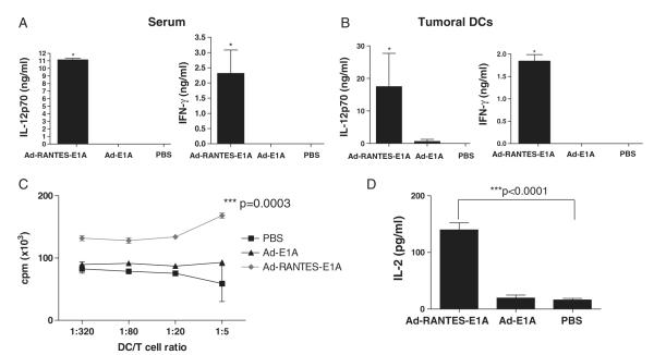
Dendritic cells (DCs) are professional antigen (Ag)-presenting cells capable of inducing immune responses to tumor Ags and, therefore, play a central role in the induction of antitumor immunity. There is a large amount of evidence, however, about paucity of tumor-associated DCs and that DCs' immunogenic functions are suppressed in a tumor environment. Here we describe a potent in situ vaccine targeting tumoral DCs in vivo. This vaccine comprised of an oncolytic adenovirus expressing RANTES (regulated upon activation, normally T expressed, and presumably secreted) (Ad-RANTES-E1A), enhanced tumor infiltration, and maturation of Ag-presenting cells in vivo. In this study, we show that intratumoral vaccinations with Ad-RANTES-E1A induced significant primary tumor growth regression and blocked metastasis formation in JC and E.G-7 murine tumor models. This vaccine recruited DCs, macrophages, natural killer cells, and CD8+ T cells to the tumor site, and thus enhanced Ag-specific cytotoxic T lymphocyte responses and natural killer cell responses. DCs purified from the Ad-RANTES-E1A-treated E.G-7 tumors secreted significantly higher levels of interferon-gamma and interleukin-12, as compared with control groups and more efficiently enhanced CD8+ T-cell response. This in situ immunization strategy could be a potent antitumor immunotherapy approach for aggressive established tumors.


Figures

FIGURE 1
FIGURE 1
Construction and characterization of Ad-RANTES-E1A and Ad-E1A vectors. A, Schematic representation of pShuttle vectors is shown. IRES indicates internal ribosome entry site; mRANTES, mouse regulated upon activation, normally T expressed, and presumably secreted. B, The integrity of the RANTES, IRES, and E1A in the produced adenovirus was verified by PCR on viral DNA template. Lane 1, PCR product of mRANTES (276 bp); lane 2, PCR product for IRES (610 bp); lane 3, PCR product for E1A (2100 bp); lanes 4 and 5, 100 bp ladder and 1 kb ladder (New England Biolabs), respectively. C, In vitro expression of RANTES. Western blot analysis of cell extracts from JC cells infected with Ad-RANTES-E1A (lane 1) and Ad-E1A (lane 2). Western blots were performed as described in Materials and Methods. Molecular weight in kDa is indicated on the right and the RANTES band (~7.9 kDa) is indicated by the arrow on the left. D, Secretion of RANTES from the transduced cells by the recombinant adenoviruses was quantitated by ELISA in the supernatants of 293 HEK and JC tumor cells. ELISA indicates enzyme-linked immunosorbent assay; HEK, human embryonic kidney cell; PCR, polymerase chain reaction.
FIGURE 2
FIGURE 2
RANTES expression in the tumor site attracts the antigen-presenting cells into the tumor mass. A, 5 × 10⁵ JC tumor cells were subcutaneously inoculated and the established tumors were injected with 1010 i.f.u. of Ad-RANTES-E1A, Ad-E1A, or PBS when they reached 5 to 7 mm in diameter. Tumors were established in 3 mice for each treatment group and processed separately. Forty-eight hours later, tumors were resected and total tumor single-cell suspensions (side and forward) scatter analyzed by flow cytometry depicted on left panel, APCs (gated as R1) were stained with antimouse CD11c, CD11b, and CD80 mAbs (right panel, flow cytometry results for CD11c and CD11b expression). DCs were gated as CD11c+ CD11b+ cells (gate R2); macrophages were gated as CD11c CD11b+ (gate R3). Mean percentage of DCs (B, upper panel). Maturation status of DCs was determined by staining with monoclonal antibodies to mouse CD80 and CD40 [B (lower panel) and C]. D, Percentage of tumor infiltrating CD8+ T cells. Data presented for 3 mice. DC indicates dendritic cell; mAbs, monoclonal antibodies; PBS, phosphate buffered saline; RANTES, regulated upon activation, normally T expressed, and presumably secreted.
FIGURE 3
FIGURE 3
Treatment with Ad-RANTES-E1A eradicates established tumors and protects from metastasis. A, 5 × 10⁵ JC cells were injected subcutaneously into the right flank of Balb/c mice. Groups of mice (N = 6) were injected intratumorally with 1 × 10¹⁰ i.f.u. of Ad-RANTES-E1A, Ad-E1A, or PBS on days 7, 8, and 14 after JC cell inoculation. Tumors were resected when they reached about 10 mm in diameter in control group (day 15 to 16 after the first adenoviral treatment). Error bars represent SEM. B, Mice were euthanized on day 21 after the first adenoviral injection. Organs from 3 mice of each group were collected and fixed in Bouin’s solution. Hematoxylin-eosin staining was performed on formalin-fixed paraffin-embedded lung and liver tissues from mice bearing JC breast tumors. Representative pictures of 9 sections (3 sections, 5-μm thick from 3 different mice of each group) at 20 × magnification. Upper panel, Liver sections; lower panel, lung sections. Arrows indicate JC metastatic tumor nodules. C, Parental E.G7 tumors were established SC into the right flank. Graph shows tumor growth in mice intratumorally injected with Ad-RANTES-E1A, Ad-E1A, and PBS as described above. D, Mice were inoculated with E.G-7 tumor cells (distant tumors) subcutaneously into the left flank before first treatment with adenovirus. Tumors were resected when they reached about 10 mm in diameter in PBS group (day 15 to 16 after the first adenoviral treatment). Error bars represent SE. PBS indicates phosphate buffered saline; RANTES, regulated upon activation, normally T expressed, and presumably secreted.
FIGURE 4
FIGURE 4
Expansion of antigen-specific CTLs in Ad-RANTES-E1A–treated mice. A, Peripheral blood was collected from the E.G-7 mice 10 days after the final adenoviral immunization. Left panel, Peripheral blood lymphocytes were stained with CD8-FITC and SIINFEKL-H2-Kb tetramer. The percentage of double-positive cells is indicated in the quadrants. Right panel, Percentage of tetramer-positive CD8+ T cells in the peripheral blood of 3 mice in each group. *P<0.05 between tetramer-positive CD8 T cells in the blood of Ad-RANTES-E1A–treated group compared with Ad-E1A and PBS groups. B, Spleens were harvested from the JC-bearing mice 10 days after final immunization and frequency of IFN-γ secreting T cells in response to JC lysates was measured by EliSpot assay. Millipore MultiScreen-HA plates were coated with antimouse IFN-γ Ab. Splenocytes (105 cells/well) were cultured for 20 hours at 37°C in a 5% CO2 incubator in the complete medium with tumor lysates of JC. C, Total splenocytes from E.G-7–bearing mice were prepared 10 days after the last immunization and cocultured with 10 μg/ml of OT-I peptide for 20 hours. The frequency of SIINFEKL-specific T cells was determined by EliSpot assay. The results were evaluated with an automated EliSpot reader system. The number of IFN-γ–producing lymphocytes was evaluated in triplicate wells. Average ± SD for each mouse was calculated and the assays were repeated twice. Ab indicates antibody; CTL, cytotoxic T lymphocyte; EliSpot, enzyme-linked immunosorbent spot; FITC, fluorescein isothiocyanate; IFN, interferon; PBS, phosphate buffered saline; RANTES, regulated upon activation, normally T expressed, and presumably secreted.
FIGURE 5
FIGURE 5
CTL and NK cell responses were induced by Ad-RANTES-E1A intratumoral vaccination in JC and E.G-7 tumor models. Spleens were harvested from tumor-bearing mice. Splenocytes were tested for cytolytic activity in a standard 6-hour 51Cr-release assay. A, Upper panel, CTLs (1.5 × 10⁶ cells) were stimulated with JC tumor lysates at a T cell:JC tumor cell ratio of 2:1 for 5 days. Target cells labeled with 51Cr were placed in each well of 96-well plates, and 50 μL of effector T cells for each dilution was added. The supernatant from each well was harvested, and the amount of 51Cr radioactivity released was measured in a γ counter. Lower panel, JC-specific CTL response in Ad-RANTES-E1A–treated mice. 51Cr-labeled CT26 colon carcinoma cells were used as syngeneic negative control. About 2 μg of monoclonal antimouse CD8 mAbs were added for the blocking assay; excess of Yac-1 cells was added to block NK cell activity (negative control). E:T ratio is 100:1. B, Spleens were harvested from E.G-7–bearing mice. Upper panel, CTLs were stimulated with 10 μg/mL of OT-I peptide for 5 days. E.G-7 target cells labeled with 51Cr were placed in each well of 96-well plates, and 50 μl of effector T cells for each dilution was added. The supernatant from each well was harvested, and the amount of 51Cr radioactivity released was measured in a γ counter. Lower panel, E.G-7–specific CTL response in Ad-RANTES-E1A–treated mice. 51Cr-labeled EL-4 cells were used as negative control. About 2 μg of monoclonal antimouse CD8 mAbs were added for the blocking assay; excess of Yac-1 cells was added to block NK cell activity; E:T ratio is 100:1. The assays were performed 3 times or more. C. Total spleen (upper panel) and tumor (lower panel) cell suspensions were stained with antimouse DX5+ Ab and percentage of DX5+ NK cells was evaluated by flow cytometry in 3 mice of each group. D, NK functional response was induced by Ad-RANTES-E1A intratumoral vaccination. Upper panel, splenocytes from Ad-RANTES-E1A, Ad-E1A, or PBS-treated mice were purified and cultured overnight in complete medium with 20 U/mL of IL-2. About 2 × 10⁵ splenocytes were plated in 96-well plates and incubated with 51Cr labeled Yac-1 cells. Lower panel, NK cells from Ad-RANTES E1A mice were cocultured with 51Cr-labeled CT26 (negative control) and with antimouse NK blocking Ab (PK 136) for 6 hours at 37°C and 5% CO2. The assays were performed 3 times in triplicates. CTL indicates cytotoxic T lymphocyte; E:T, effector:target; IL, interleukin; mAbs, monoclonal antibodies; NK, natural killer; PBS, phosphate buffered saline; RANTES, regulated upon activation, normally T expressed, and presumably secreted.
FIGURE 6
FIGURE 6
Elevated levels of IL-12 and IFN-γ in tumor infiltrating DCs and blood serum of Ad-RANTES-E1A–treated mice and high immunostimulatory function of tumor-infiltrating DCs. A, Blood serum was collected from 3 mice of each group (Ad-RANTES-E1A, Ad-E1A, and PBS) 5 days after the last adenoviral treatment. Levels of IL-12 and IFN-γ were measured by ELISA. B, Tumors were established subcutaneously into the right and left flanks of 6 mice of each treatment group. Forty-eight hours after the intratumoral adenoviral injections, tumors were resected and tumoral DCs were purified as described in Materials and Methods. Purified DCs were cultured overnight in a 96-well plate in triplicates and level of the secreted cytokines in DCs-conditioned medium was measured by ELISA. *P<0.05 between levels in Ad-RANTES-E1A group compared with Ad-E1A and PBS groups. C and D, E.G-7 tumors were established subcutaneously into the right and left flanks of 6 mice of each treatment group. Forty-eight hours after the intratumoral adenoviral injections, tumors were resected and tumoral DCs were purified as described in Materials and Methods. Graded numbers of CD11c+-enriched tumoral DCs cultured for 3 days in different culture were cocultured with 3 × 10⁵ purified naive CD8+ T cells in the presence of anti-CD3 for 3 days. In the last 16 hours, 3H-thymidine (1 μCi/well) was added into the cell culture to monitor the T-cell proliferation (C) and IL-2 secretion (D) was measured 16 hours later. Results shown are representative of 3 different experiments. ***P<0.0001 and *P<0.05 for cytokine secretion and proliferation primed by tumoral DCs of Ad-RANTES-E1A–treated group compared with Ad-E1A and PBS groups. DC indicates dendritic cell; ELISA, enzyme-linked immunosorbent assay; IFN, interferon; IL, interleukin; PBS, phosphate buffered saline; RANTES, regulated upon activation, normally T expressed, and presumably secreted.

Similar articles

Cited by

References

    1. Banchereau J, Steinman RM. Dendritic cells and the control of immunity. Nature. 1998;392:245. - PubMed
    1. Steinman RM. The control of immunity and tolerance by dendritic cell. Pathol Biol (Paris) 2003;51:59. - PubMed
    1. Shortman K, Liu YJ. Mouse and human dendritic cell subtypes. Nat Rev Immunol. 2002;2:151. - PubMed
    1. Godelaine D, Carrasco J, Lucas S, et al. Polyclonal CTL responses observed in melanoma patients vaccinated with dendritic cells pulsed with a MAGE-3A1 peptide. J Immunol. 2003;171:4893. - PubMed
    1. Kugler A, Stuhler G, Walden P, et al. Regression of human metastatic renal cell carcinoma after vaccination with tumor cell-dendritic cell hybrids. Nat Med. 2000;6:332. - PubMed

Publication types

MeSH terms