. 2009;4(2):e4579.
doi: 10.1371/journal.pone.0004579. Epub 2009 Feb 24.

Localisation and function of the endocannabinoid system in the human ovary

Affiliations

Localisation and function of the endocannabinoid system in the human ovary

Mona R El-Talatini et al. PLoS One. 2009.

Abstract

Background: Although anandamide (AEA) had been measured in human follicular fluid and is suggested to play a role in ovarian follicle and oocyte maturity, its exact source and role in the human ovary remains unclear.

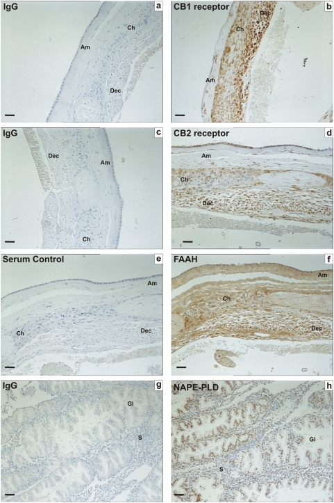
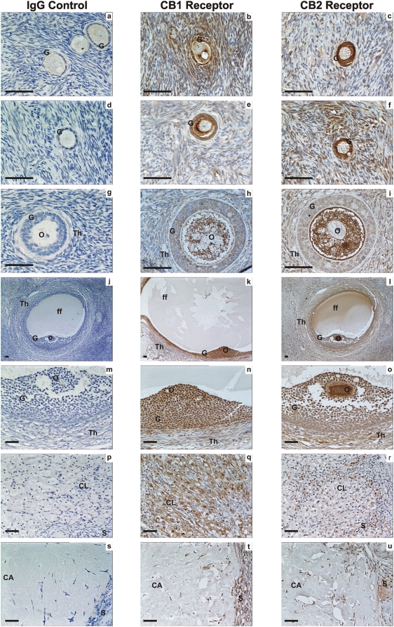
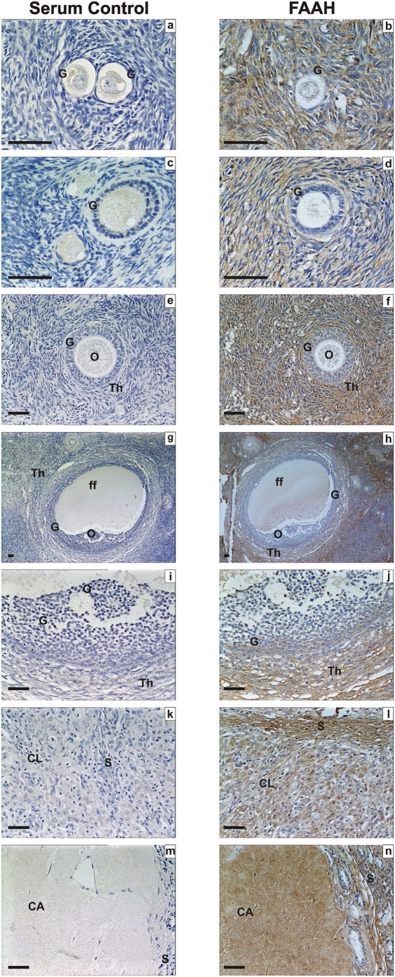
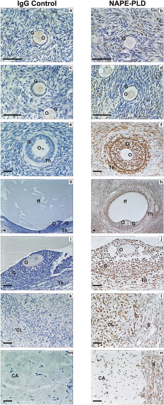
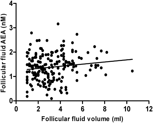
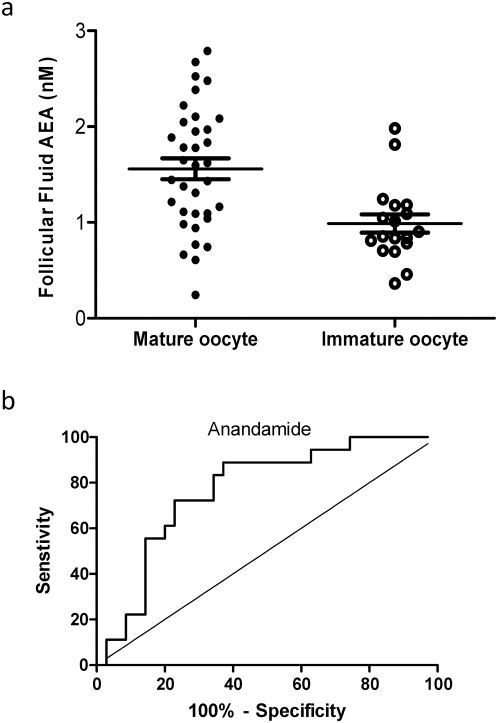
Methods and findings: Immunohistochemical examination of normal human ovaries indicated that the endocannabinoid system was present and widely expressed in the ovarian medulla and cortex with more intense cannabinoid receptor 2 (CB2) than CB1 immunoreactivity in the granulosa cells of primordial, primary, secondary, tertiary follicles, corpus luteum and corpus albicans. The enzymes, fatty acid amide hydrolase (FAAH) and N-acyclphosphatidylethanolamine-phospholipase D (NAPE-PLD), were only found in growing secondary and tertiary follicles and corpora lutea and albicantes. The follicular fluid (FF) AEA concentrations of 260 FF samples, taken from 37 infertile women undergoing controlled ovarian hyperstimulation for in vitro fertilisation and intracytoplasmic sperm injection with embryo transfer, were correlated with ovarian follicle size (P = 0.03). Significantly higher FF AEA concentrations were also observed in mature follicles (1.43+/-0.04 nM; mean+/-SEM) compared to immature follicles (1.26+/-0.06 nM), P = 0.0142 and from follicles containing morphologically assessed mature oocytes (1.56+/-0.11 nM) compared to that containing immature oocytes (0.99+/-0.09 nM), P = 0.0011. ROC analysis indicated that a FF AEA level of 1.09 nM could discriminate between mature and immature oocytes with 72.2% sensitivity and 77.14% specificity, whilst plasma AEA levels and FF AEA levels on oocyte retrieval day were not significantly different (P = 0.23).

Conclusions: These data suggest that AEA is produced in the ovary, is under hormonal control and plays a role in folliculogenesis, preovulatory follicle maturation, oocyte maturity and ovulation.


Conflict of interest statement

Competing Interests: The authors have declared that no competing interests exist.

Figures

Figure 1
Figure 1. Immunohistochemical staining for CB1, CB2, FAAH and NAPE-PLD on control tissues.
Images on the left side of the panel represent the negative control (IgG/serum) and images on the right side of the panel are sections incubated with specific antibodies/antisera. The tissues used in a–f are human fetal membranes whereas g and h are human endometrium. CB1, CB2 and FAAH Immunoreactivity was observed in the amnion (Am), chorion (Ch) and decidua (Dec) of term fetal membranes, whilst NAPE-PLD immunoreactivity was observed mainly in the endometrial glands (Gl) with sparse staining in endometrial stroma (S). The images are representatives from at least six samples and were taken at 200× magnification. Bar = 50 µm.
Figure 2
Figure 2. Immunohistochemical staining for CB1 and CB2 receptors.
Images on the left side of the panel represent the negative control (IgG) and images in the middle are for CB1 on the right are for CB2. Images a, b and c are primordial follicle; d, e and f are primary follicles; g, h and i are secondary follicles; j, k and l are low power images of tertiary follicles; m, n an o are high power images of tertiary follicles, p, q and r are corpus luteum, r, s and t are images of the corpus albicans. Granulosa cells (G), theca cell layers (Th), the oocyte (O) and follicular fluid (ff) demonstrated CB1 and CB2 immunoreactivity as did lutein-granulosa cells of the corpus luteum (CL) and corpus albicans (CL), and septa (S) between lobes of these postovulatory bodies. The images are representatives from at least two structures and taken at 50×, 200× or 400× magnification. Bar = 50 µm. Ovarian follicles were classified as primordial, primary, secondary, tertiary, corpus luteum, and corpus albicans .
Figure 3
Figure 3. Immunohistochemical staining for FAAH.
Images on the left side of the panel represent the negative control (non-immune rabbit serum and images on the right are for FAAH. Images a and b are primordial follicle; c and d are primary follicles; e and f are secondary follicles; g and h are low power images of tertiary follicles; i and j are high power images of tertiary follicles, k and l are corpus luteum, and m and n are images of the corpus albicans. Granulosa cells (G), theca cell layers (Th), the oocyte (O) and follicular fluid (ff) demonstrated FAAH immunoreactivity as did lutein-granulosa cells of the corpus luteum (CL) and corpus albicans (CL), and septa (S). There was a gradation of staining in the granulosa cells of the tertiary follicle with greatest intensity in the mural cells (panel j). The images are representatives from at least two structures and taken at 50×, 200× or 400× magnification. Bar = 50 µm.
Figure 4
Figure 4. Immunohistochemical staining for NAPE-PLD.
Images in the left side of the panel represent the negative control (rabbit IgG) and images on the right are for NAPE-PLD. Images a, and b are primordial follicles, c and d are primary follicles, e and f are secondary follicles; g and h are low power images of tertiary follicles, i and j are high power images of tertiary follicles, k and i are images of corpus luteum, and m and n are images of corpus albicans. Granulosa cells (G), theca cell layers (Th), the oocyte (O) and follicular fluid (ff) demonstrated NAPE-PLD immunoreactivity as did lutein-granulosa cells of the corpus luteum (CL) and corpus albicans (CL) and septa (S). The images are representative from at least two structures and taken at 50×, 200×, or 400× magnification. Bar = 50 µm.
Figure 5
Figure 5. Spearman correlation between follicular fluid AEA concentration and follicle size.
A positive correlation between the size of preovulatory follicle (in ml) and the concentrations of follicular fluids AEA from IVF/ICSI patients is shown. Follicular fluids from 193 follicles were analysed, R = 0.1304; P = 0.03.
Figure 6
Figure 6. Prediction of oocyte maturity from follicular fluid AEA measurements.
Panel a shows the follicular fluid AEA concentrations from follicles that gave rise to mature and immature oocytes during ICSI cycles. The long horizontal bar represents the mean and the shorter bars the sem. Panel b shows a receiver-operating characteristic curve (ROC) analysis for the prediction of the production of mature oocytes from follicular AEA concentration. The sensitivity and specificity relationship for measurements of anandamide in follicular fluid is plotted. The optimum cut off point for the identification of mature oocytes at 1.09 nM, provided a sensitivity of 72.2%, (95CI = 46.52% to 90.31%) and a specificity of 77.14% (95CI = 59.86 to 89.58%) and likelihood ratio of 3.16. The area under the curve was 0.768±0.067, (Mean±sem; 95%CI = 0.63–0.89, P = 0.001).

References

    1. Devane WA, Hanus L, Breuer A, Pertwee RG, Stevenson LA, et al. Isolation and structure of a brain constituent that binds to the cannabinoid receptor. Science. 1992;258:1946–1949. - PubMed
    1. Childers SR, Breivogel CS. Cannabis and endogenous cannabinoid systems. Drug and Alcohol Dependence. 1998;51:173–187. - PubMed
    1. Habayeb OM, Bell SC, Konje JC. Endogenous cannabinoids: Metabolism and their role in reproduction. Life Sciences. 2002;70:1963–1977. - PubMed
    1. Piomelli D, Giuffrida A, Calignano A, Rodriguez de Fonseca F. The endocannabinoid system as a target for therapeutic drugs. Trends in Pharmacological Sciences. 2000;21:218–224. - PubMed
    1. Galiegue S, Mary S, Marchand J, Dussossoy D, Carriere D, et al. Expression of central and peripheral cannabinoid receptors in human immune tissues and leukocyte subpopulations. European Journal of Biochemistry. 1995;232:54–61. - PubMed

Publication types