Comparative Study
. 2009 Apr 21;48(15):3269-78.
doi: 10.1021/bi8023493.

Evidence for an active role of IF3mt in the initiation of translation in mammalian mitochondria

Affiliations
Comparative Study

Evidence for an active role of IF3mt in the initiation of translation in mammalian mitochondria

Brooke E Christian et al. Biochemistry. .

Abstract

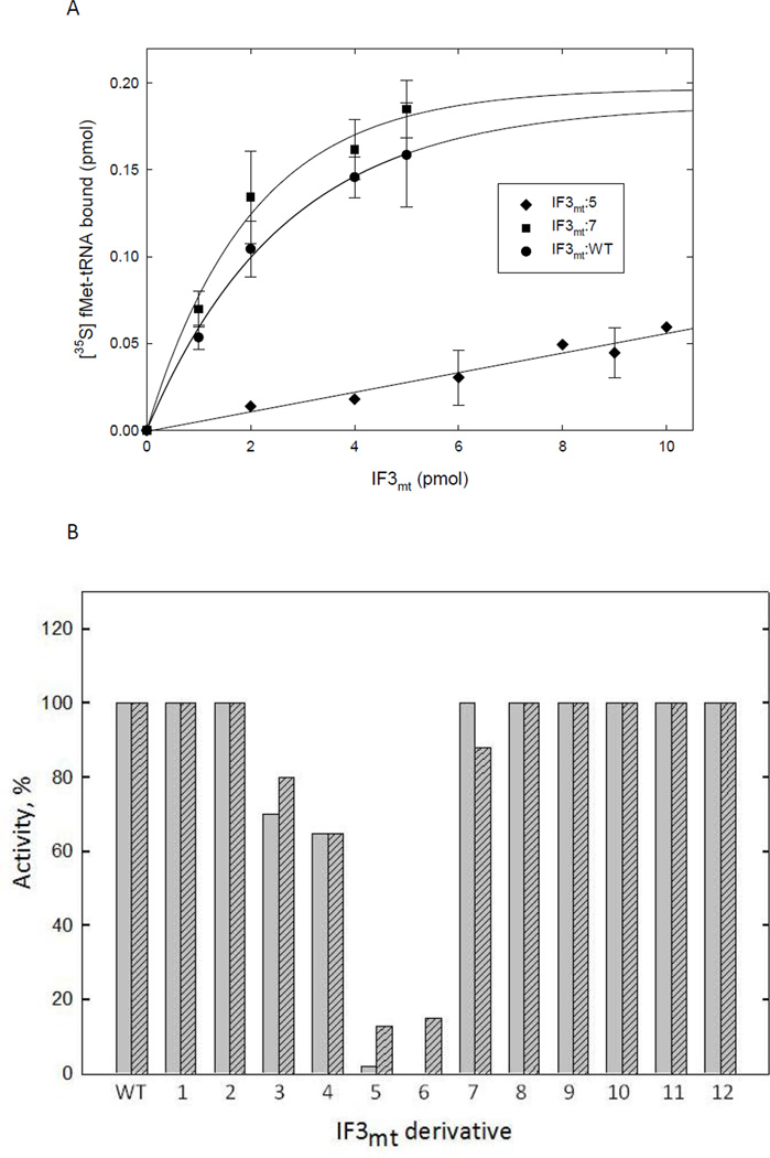
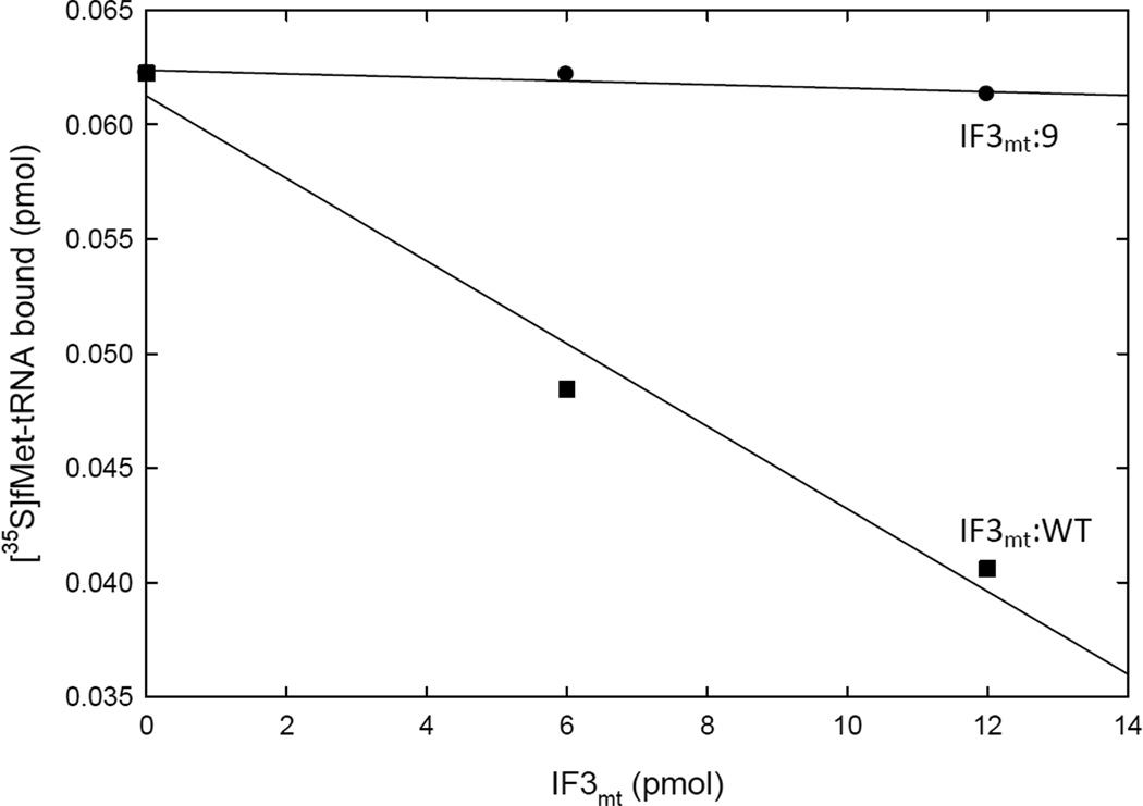
Mitochondrial translational initiation factor 3 (IF3(mt)) is a 29 kDa protein that has N- and C-terminal domains, homologous to prokaryotic IF3, connected by a linker region. The homology domains are preceded and followed by short extensions. No information is currently available on the specific residues in IF3(mt) important for its activity. On the basis of homology models of IF3(mt), mutations were designed in the N-terminal, C-terminal, and linker domains to identify the functionally important regions. Mutation of residues 170-171, and 175 in the C-terminal domain to alanine resulted in a nearly complete loss of activity in initiation complex formation and in the dissociation of mitochondrial 55S ribosomes. However, these mutated proteins bind to the small (28S) subunit of the mammalian mitochondrial ribosome with K(d) values similar to that of the wild-type factor. These mutations appear to lead to a factor defective in the ability to displace the large (39S) subunit of the ribosome from the 55S monosomes in an active process. Other mutations in the N-terminal domain, the linker region, and the C-terminal domain had little or no effect on the ability of IF3(mt) to promote initiation complex formation on mitochondrial 55S ribosomes. Mutation of residues 247 and 248 in the C-terminal extension abolished the ability of IF3(mt) to reduce the level of binding of fMet-tRNA to the ribosome in the absence of mRNA. Our results suggest that IF3(mt) plays an active role in initiation of translation.


Figures

Figure 1
Figure 1
Domain organization and model of IF3mt. A. Schematic representation of E. coli IF3 and IF3mt showing the N- and C-terminal homology domains and the linker regions. IF3mt has additional N- and C-terminal extensions not present in the E. coli factor. The leader specifies mitochondrial import and is not present in the constructs used here. B. The 3-D model of IF3mt prepared using Insight II. The N-terminal domain was modeled after the crystal structure of the N-terminal domain of B. stearothermophilus IF3 (PDB coordinates 1TIF (26)), and the C-domain was modeled after the NMR structure of the mouse IF3mt (PDB coordinates 2CRQ, unpublished). The N- and C-terminal extensions are not shown and are predicted to be disordered.
Figure 2
Figure 2
Mutations in IF3mt. A. Conservation of the charged residues mutated in IF3mt:1–8. Positively charged residues are shown in dark gray and negatively charged residues are shown in light gray. The numbers above each cluster of residues indicates the IF3mt derivative containing mutations of those residues. B. stearothermophilus IF3 does not have residues corresponding to IF3mt:1. These residues are present in E. coli IF3 as KRVQT. B. Sequence conservation of IF3mt:9–12 among the vertebrate lineage. Positively charged residues are shown in dark gray and negatively charged residues are shown in light gray. C. Model of IF3mt showing IF3mt:1–8. The N-terminal domain is shown in red, the linker is in green, and the mutated residues are in blue. IF3mt:1 could not be modeled based on the crystal structure of B. stearothermophilus because these residues are not present in this IF3 nor are they present in the NMR structure of the N-domain of E. coli IF3 (38). IF3mt:1 has been placed in the model at the N-terminus of the protein for illustrative purposes. The C-terminal homology domain is shown in blue and the mutated residues are shown in orange. Part of the C-terminal extension is shown in yellow.
Figure 3
Figure 3
Effect of mutated derivatives of IF3mt on initiation complex formation. A. Effect of IF3mt:WT, IF3mt:7, and IF3mt:5 on initiation complex formation on mitochondrial 55S ribosomes. [35S]fMet-tRNA binding to mitochondrial 55S particles was tested in the presence of saturating amounts of IF2mt using IF3mt:WT (●), IF3mt:7 (■), or IF3mt:5 (◆). A blank representing the amount of [35S]fMet-tRNA bound to ribosomes in the absence of IF3mt (~0.1 pmol) was subtracted from each value. B. Summary of the activities of IF3mt:1–12 in initiation complex formation. This assay primarily measures ribosome dissociation. The activity of IF3mt:WT was set to 100 % and the mutated derivatives were compared to that value using the linear regions of the dose response curves. The activity on 70S ribosomes is shown in gray while activity on mitochondrial ribosomes is in striped gray. Derivatives identical to wild-type within error are shown as 100 %.
Figure 4
Figure 4
Effect of IF3mt and its mutated derivates on the dissociation of mitochondrial 55S ribosomes. Fractionation profiles of mitochondrial 55S ribosomes after centrifugation on a 10– 30 % sucrose gradient. Mitochondrial 55S ribosomes (8 pmol) were incubated as described in Materials and Methods in the absence (A) or presence of 80 pmol IF3mt (B) or its mutated derivative IF3mt:6 (C) and subsequently subjected to centrifugation on a 10–30 % sucrose density gradient. Gradients were fractionated while monitoring the A254. D. Percentage of mitochondrial 55S ribosomes remaining after the addition of IF3mt:WT or its mutated derivatives as measured by sucrose density gradient centrifugation.
Figure 5
Figure 5
Binding of IF3mt:6 to 28S subunits. Binding assays were performed using a Microcon spin column as described (18). Inset: Calibration curve of IF3mt: WT using a colorimetric assay and a dot blot apparatus. This curve was used to determine the amount of IF3mt bound to mitochondrial 28S ribosomes in the Microcon centrifugation assay as described in Materials and Methods.
Figure 6
Figure 6
IF3mt-mediated inhibition of fMet-tRNA binding to mitochondrial 28S subunits in the absence of mRNA. Mitochondrial 28S subunits were incubated with saturating amounts of IF2mt and [35S]fMet-tRNA in the presence or absence of IF3mt:WT or its C-terminal extension mutated derivative IF3mt:9 as described (16). The amount of [35S]fMet-tRNA remaining bound to the filter in the presence of IF3mt:WT (■) or IF3mt:9 (●) was determined using a nitrocellulose filter binding assay.
Figure 7
Figure 7
Effects of mutations in the C-terminal domain of E. coli IF3 and IF3mt. A. Mutated residues in the C-terminal domain of E. coli IF3 and their effects on activity in initiation complex formation on E. coli ribosomes. The activity remaining was based on 70S dissociation, one of several assays carried out with these mutated derivatives of E. coli IF3. The most drastic effects on activity are seen with the mutation in the center of the α-helix (Helix 4) on the back of the protein. B. Mutated residues in the C-terminal domain of IF3mt and the effects of these mutations on activity in initiation complex formation on mitochondrial ribosomes. The most drastic effects are seen with the two mutations located at the base of Helix 3.
Figure 8
Figure 8
Models for the mechanism of IF3mt in the dissociation of mitochondrial ribosomes. A. In the passive model, the subunits are in equilibrium with the 55S monosome (passive step 1). IF3mt binds to free 28S subunits, preventing reassociation with the 39S subunit (passive step 2). B. In the active model, IF3mt interacts with the 55S particle, forming a transient 28S:IF3mt:39S complex (active step 1), which then dissociates into a 28S:IF3mt complex and free 39S subunits (active step 2).

Similar articles

Cited by

References

    1. Lin MT, Beal MF. Mitochondrial dysfunction and oxidative stress in neurodegenerative diseases. Nature. 2006;443:787–795. - PubMed
    1. Scheffler I. Mitochondria. New York: Wiley-Liss, Inc; 1999.
    1. Sharma MR, Koc EC, Datta PP, Booth TM, Spremulli LL, Agrawal RK. Structure of the mammalian mitochondrial ribosome reveals an expanded functional role for its component proteins. Cell. 2003;115:97–108. - PubMed
    1. Gualerzi C, Pon C. Initiation of mRNA translation in prokaryotes. Biochemistry. 1990;29:5881–5889. - PubMed
    1. Cummings H, Hershey J. Translation initiation factor IF-1 is essential for cell viability in Escherichia coli . J. Bacteriol. 1994;176:198–205. - PMC - PubMed

Publication types

MeSH terms