Review
. 2009 Mar 31;48(12):2574-85.
doi: 10.1021/bi900108v.

Prion diseases and their biochemical mechanisms

Affiliations
Review

Prion diseases and their biochemical mechanisms

Nathan J Cobb et al. Biochemistry. .

Abstract

Prion diseases, also known as the transmissible spongiform encephalopathies (TSEs), are a group of fatal neurodegenerative disorders that affect humans and animals. These diseases are intimately associated with conformational conversion of the cellular prion protein, PrP(C), into an oligomeric beta-sheet-rich form, PrP(Sc). A growing number of observations support the once heretical hypothesis that transmission of TSE diseases does not require nucleic acids and that PrP(Sc) alone can act as an infectious agent. The view that misfolded proteins can be infectious is also supported by recent findings regarding prion phenomena in yeast and other fungi. One of the most intriguing facets of prions is their ability to form different strains, leading to distinct phenotypes of TSE diseases. Within the context of the "protein-only" model, prion strains are believed to be encoded in distinct conformations of misfolded prion protein aggregates. In this review, we describe recent advances in biochemical aspects of prion research, with a special focus on the mechanism of conversion of prion protein to the pathogenic form(s), the emerging structural knowledge of fungal and mammalian prions, and our rapidly growing understanding of the molecular basis of prion strains and their relation to barriers of interspecies transmissibility.


Figures

Figure 1
Figure 1
Schematic representation of the potential mechanism of neuroinvasion in transmissible spongiform encephalopathies. (A) Inital uptake of the TSE agent from the intestinal lumen has been proposed to occur through a number of alternative mechanisms, including M cell transcytosis (i), ferritin-dependent trancytosis through intestinal epithelial cells (ii), or via direct capture by dendritic cells (iii). While phagocytic cells such as macrophages appear to degrade PrPSc (iv), dendritic cells may deliver the TSE agent to follicular dendritic cells (FDCs) where early accumulation of PrPSc occurs (v). (B) After amplification of the TSE agent in lymphoid tissue such as the GALT and spleen, invasion of the nervous system is believed to proceed through peripheral nerves. Retrograde transport of the TSE agent is believed to occur along two distinct pathways, following efferent fibers of the sympathetic and parasympathetic nerves to the CNS. (C) Retrograde transport and propagation of PrPSc along neuronal processes may occur by step-wise interactions along the cell surface (ia, ib), via extracellular deposits (ii), or by vesicle-mediated mechanisms (iiia, iiib). See text for details
Figure 2
Figure 2
Comparison of structural models for fungal and mammalian prion protein aggregates. (A) Structural models of yeast fungal prion protein amyloids. (i) X-ray diffraction structure of microcrystals formed by the peptide GNNQQNY, corresponding to a fragment of yeast prion protein Sup35 (adapted from ref , reprinted by permission from Macmillan Publishers Ltd.). The peptides form parallel and in-register β-sheets which associate at a dry interface known as the ‘steric zipper’. (ii) Left-handed β-solenoid structure determined by solid-state NMR for amyloid fibrils formed by yeast protein protein HET-s, where each molecule winds to form two three-stranded layers of the amyloid core (adapted from ref , reprinted with permission from AAAS). (iii) A general model for amyloid fibrils formed by yeast prion proteins Sup35, Ure2p, and Rnq1 (adapted from ref , Copyright (2008) John Wiley and Sons, Inc.). In all cases, solid-state NMR has revealed a parallel and in-register packing of individual molecules to form single layers where same residues are perfectly aligned with their counterparts on neighboring molecules. (B) Structural models of mammalian PrPSc. (i) The β-helical model, where residues ~90-175 are shown to form left-handed β-helicies that associate into trimers, leaving the most C-terminal helices of monomeric PrPC intact (adapted from ref , Copyright (2004) National Academy of Sciences, U.S.A.). (ii) The spiral model of PrPSc depicts the amyloid core as being comprised of a three stranded β-sheet and isolated β-strand, with complete retention of all three native α-helicies (adapted from ref , Copyright (2004) National Academy of Sciences, U.S.A.). (iii) Parallel and in-register β-structure model determined experimentally for recombinant PrP amyloid fibrils (adapted from ref , Copyright (2007) National Academy of Sciences, U.S.A.). In this model, residues ~160–220 form the PrP amyloid core (native disulfide bond shown in green), with tight interdigitation of side chains. Individual monomers stack to form single molecule layers so that same residues are perfectly aligned. In all cases, arrows indicate the long fibrillar axis.
Figure 3
Figure 3
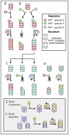
Illustration of the phenomenon of prion strains and transmissibility barriers. Different PrPC sequences (differentiated by color) dictate the spectrum of allowable PrPSc conformations (depicted as different shapes), and these conformations represent different prion strains. For simplicity, transmission barriers are depicted as absolute, although in reality such barriers are often characterized by prolonged incubation time. (A) Infection of species X with a specific prion strain derived from the same species results in faithful propagation of strain characteristics. (B) Passaging of the same prion strain from species X to species Y and Z (which express non-homologous PrPC) may have different outcomes. If the PrPSc conformation of the donor strain from species X is not accessible to PrPC of the host species, a barrier to transmission is observed as illustrated for species Y. On the other hand, if the conformation of the donor strain is accessible to the host PrPC, transmission occurs, resulting in emergence of a new strain of prion in the host species (as illustrated by species Z). (C) The newly formed species Z prion strain oftentimes displays species-specific transmissibility characteristics similar to those of the original template. (D) Other species Z prion strains may, however, show transmission barriers that are distinct from those observed for the specific template-adapted strain shown in Panel C. Thus, infectivity is associated with conformational properties of a particular prion strain. (E and F) Models of ‘strain-switching’, a phenomenon that may occur upon cross-species transmission of a specific prion strain. (E) Strain conversion model: PrPC substrate adopts a conformation that is not identical to that of a non-homologous PrPSc template. (F) Strain selection model: a disease phenotype (strain) is associated with multiple PrPSc conformers, one of which is ‘dominant’ in a particular species. Upon cross-species transmission, non-homologous host PrPC selects the PrPSc template most compatible with its amino acid sequence. In either case, new PrPSc conformations associated with the emerging strain may confer distinct transmission barriers and disease phenotype.

References

    1. Prusiner SB. Novel proteinaceous infectious particles cause scrapie. Science. 1982;216:136–144. - PubMed
    1. Prusiner SB. Prions. Proc Natl Acad Sci U S A. 1998;95:13363–13383. - PMC - PubMed
    1. Caughey B, Chesebro B. Transmissible spongiform encephalopathies and prion protein interconversions. Adv Virus Res. 2001;56:277–311. - PubMed
    1. Collinge J. Prion diseases of humans and animals: their causes and molecular basis. Annu Rev Neurosci. 2001;24:519–550. - PubMed
    1. Aguzzi A, Polymenidou M. Mammalian prion biology: one century of evolving concepts. Cell. 2004;116:313–327. - PubMed

Publication types