Successful management of multiple permanent pacemaker complications--infection, 13 year old silent lead perforation and exteriorisation following failed percutaneous extraction, superior vena cava obstruction, tricuspid valve endocarditis, pulmonary embolism and prosthetic tricuspid valve thrombosis
- PMID: 19239701
- PMCID: PMC2649923
- DOI: 10.1186/1749-8090-4-12
Successful management of multiple permanent pacemaker complications--infection, 13 year old silent lead perforation and exteriorisation following failed percutaneous extraction, superior vena cava obstruction, tricuspid valve endocarditis, pulmonary embolism and prosthetic tricuspid valve thrombosis
Abstract
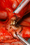
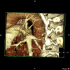
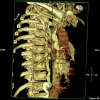
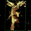
A 59 year old man underwent mechanical tricuspid valve replacement and removal of pacemaker generator along with 4 pacemaker leads for pacemaker endocarditis and superior vena cava obstruction after an earlier percutaneous extraction had to be abandoned, 13 years ago, due to cardiac arrest, accompanied by silent, unsuspected right atrial perforation and exteriorisation of lead. Postoperative course was complicated by tricuspid valve thrombosis and secondary pulmonary embolism requiring TPA thrombolysis which was instantly successful. A review of literature of pacemaker endocarditis and tricuspid thrombosis along with the relevant management strategies is presented. We believe this case report is unusual on account of non operative management of right atrial lead perforation following an unsuccessful attempt at percutaneous removal of right sided infected pacemaker leads and the incidental discovery of the perforated lead 13 years later at sternotomy, presentation of pacemaker endocarditis with a massive load of vegetations along the entire pacemaker lead tract in superior vena cava, right atrial endocardium, tricuspid valve and right ventricular endocardium, leading to a functional and structural SVC obstruction, requirement of an unusually large dose of warfarin postoperatively occasioned, in all probability, by antibiotic drug interactions, presentation of tricuspid prosthetic valve thrombosis uniquely as vasovagal syncope and isolated hypoxia and near instantaneous resolution of tricuspid prosthetic valve thrombosis with Alteplase thrombolysis.
Figures


References
-
- Hayes DL, Zipes DP. Cardiac pacemakers and cardioverter-defibrillators. In: Zipes DP, Libby P, Bonow RO, Braunwald E, editor. Braunwald's Heart Disease. Philadelphia, Pennsylvania: Elsevier Saunders; 2005. p. 784.
-
- Da-Costa A, Kirkorian G, Isaaz K, Touboul P. Secondary infections after pacemaker implantation. Rev Med Interne. 2000;21:256–65. - PubMed
-
- Sharma S, Tieyjeh IM, Espinosa RE, Costello BA, Baddour LM. Pacemaker infection due to Mycobacterium fortuitum. Scand J Infec Dis. 2005;37:66–7. - PubMed
Publication types
MeSH terms
LinkOut - more resources
Full Text Sources
Medical

