

Review
doi: 10.1016/j.cell.2009.01.044.
mRNA localization: gene expression in the spatial dimension
Affiliations
- PMID: 19239891
- PMCID: PMC2819924
- DOI: 10.1016/j.cell.2009.01.044
Item in Clipboard
Review
mRNA localization: gene expression in the spatial dimension
Cell.
.
Abstract
The localization of mRNAs to subcellular compartments provides a mechanism for regulating gene expression with exquisite temporal and spatial control. Recent studies suggest that a large fraction of mRNAs localize to distinct cytoplasmic domains. In this Review, we focus on cis-acting RNA localization elements, RNA-binding proteins, and the assembly of mRNAs into granules that are transported by molecular motors along cytoskeletal elements to their final destination in the cell.
Figures
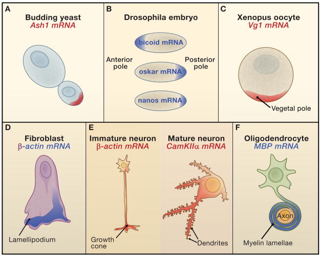
A) In budding yeast, the ASH1 mRNA localizes to the bud tip. B) In Drosophila embryos, bicoid mRNA localizes to the anterior pole; oskar and nanos mRNAs to the posterior pole. C) In Xenopus oocytes (stage IV), Vg 1 mRNA localizes to the vegetal pole. D) In chick and mammalian fibroblasts, β-actin mRNA localizes to lamellipodia. E) In developing, immature mammalian neurons, β-actin mRNA is present in distal growth cones; in mature, fully polarized pyramidal neurons, CamKIIα mRNA is present in distal dendrites. F) In mammalian oligodendrocytes, myelin basic protein (MBP) mRNA localizes to myelinating processes that ensheath neuronal axons.
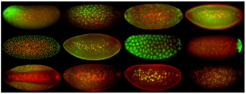
High-resolution fluorescent in situ analysis of 25% of mRNAs encoded by the Drosophila genome revealed that 71% of these display striking patterns of subcellular localization in early embryos. Some of these patterns are illustrated in this montage of photomicrographs, in which nuclei are in red and mRNAs in green. The anterior pole of the embryo is to the left, and the posterior pole to the right.
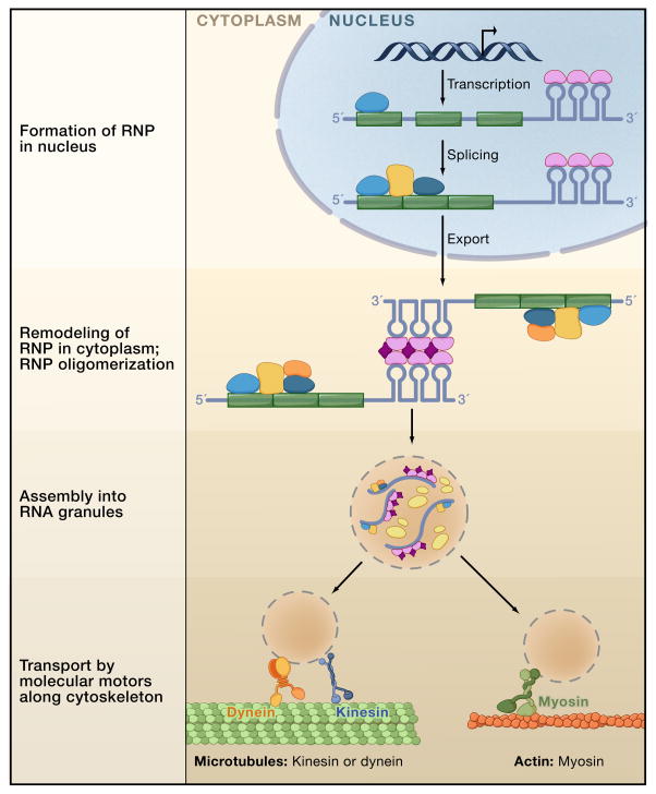
The pre-mRNA (exons in green; introns, 5′ and 3′ UTRs in grey) has cis-acting localization elements in its primary sequence. These are usually in the 3′UTR and often form stem-loop structures. RNA binding proteins (blue and purple) bind the pre-mRNA. During splicing, additional RNA binding proteins (golden and dark blue) are added to form a ribonucleoprotein (RNP) complex. Following export into the cytoplasm, the RNP is remodeled as additional proteins (orange, dark purple) are added. In some cases, the RNP can form oligomers with other RNPs through protein-protein interactions. In the cytoplasm, RNPs are assembled into RNA granules that are likely a heterogeneous population of structures containing diverse RNAs, ribosomal subunits (yellow), as well as many factors involved in translational regulation. Recent studies suggest a dynamic relationship between RNA transport granules, P-bodies and stress granules. The RNA granules associate with motor proteins and are transported by cytoskeletal elements to their final destination.
References
-
- Arn EA, Cha BJ, Theurkauf WE, Macdonald PM. Recognition of a bicoid mRNA localization signal by a protein complex containing Swallow, Nod, and RNA binding proteins. Dev Cell. 2003;4:41–51. - PubMed
Publication types
MeSH terms
Substances
Grants and funding
LinkOut - more resources
Full Text Sources
Other Literature Sources
