Review
. 2009 Feb 20;30(2):180-92.
doi: 10.1016/j.immuni.2009.01.001.

The many roles of FAS receptor signaling in the immune system

Affiliations
Review

The many roles of FAS receptor signaling in the immune system

Andreas Strasser et al. Immunity. .

Abstract

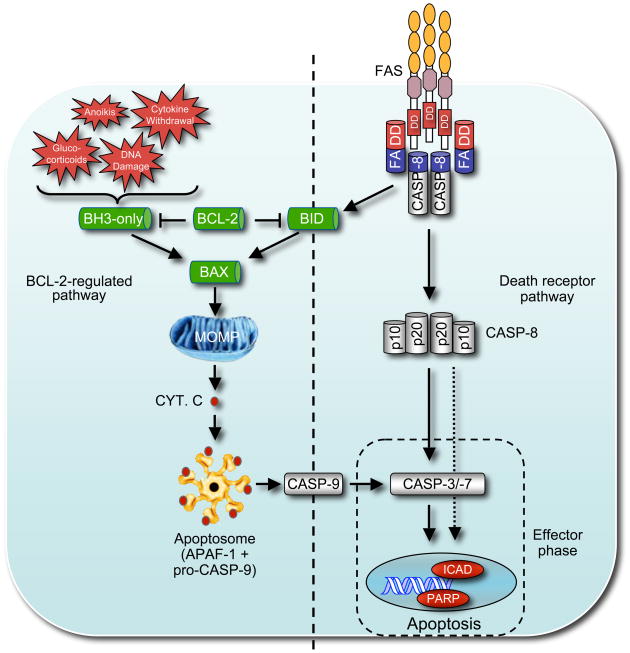
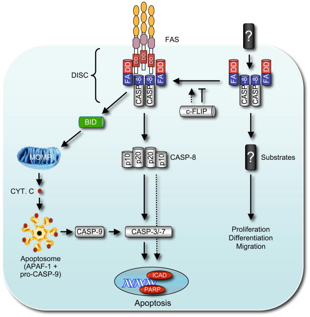
FAS belongs to the subgroup of the tumor necrosis factor receptor (TNF-R) family that contains an intracellular "death domain" and triggers apoptosis. Its physiological ligand FASL is a member of the TNF cytokine family. Studies with mutant mice and cells from human patients have shown that FAS plays critical roles in the immune system, including the killing of pathogen-infected cells and the death of obsolete and potentially dangerous lymphocytes. Fas thereby functions as a guardian against autoimmunity and tumor development. FAS triggers apoptosis through FADD-mediated recruitment and activation of caspase-8. In certain cells such as hepatocytes, albeit not lymphocytes, FAS-induced apoptosis requires amplification through proteolytic activation of the proapoptotic BCL-2 family member BID. Curiously, several components of the FAS signaling machinery have been implicated in nonapoptotic processes, including cellular activation, differentiation, and proliferation. This review describes current understanding of Fas-induced apoptosis signaling and proposes experimental strategies for future advances.


Figures

Figure 1
Figure 1
This figure shows the two distinct but ultimately converging apoptosis signaling pathways in mammals. One is activated by so-called ‘death receptors’, members of the tumor necrosis factor receptor (TNF-R) family with an intra-cellular ‘death domain’ and requires FADD-mediated activation of caspase-8 (shown on the right). Auto-processing of caspase-8 results in a hetero-tetrameric form of the protein that is able to activate the zymogens of ‘effector’ caspases, caspase-3 and caspase-7, eventually leading to cleavage of vital proteins within the cell, such as PARP or lamins, or activation of the DNAse CAD (caspase-activated DNAse) by cleavage of its inhibitor, ICAD (inhibitor of CAD). It is possible that caspase-8 may also proteolyze to a certain extent some of the classical substrates of effector caspases. The ‘BCL-2-regulated’ pathway (shown on the left) is arbitrated by the complex interplay between pro- and anti-apoptotic members of the BCL-2 protein family and involves mitochondrial outer membrane permeabilization (MOMP). MOMP results in release of cytochrome C from the inter-membrane space of the mitochondria into the cytosol to initiate apoptosome formation resulting in APAF-1-mediated activation of the ‘initiator’ caspase, caspase-9, further leading to proteolytic activation of the downstream ‘effector’ caspases, caspase-3 and caspase-7. Abbreviations: BH3-only: BCL-2 homology domain 3 only protein; MOMP: mitochondrial outer membrane permeabilization; Cyt. C: cytochrome C. ICAD: Inhibitor of caspase activated DNAse (CAD); PARP: Poly (ADP-ribose) polymerase.
Figure 2
Figure 2
This figure shows the mechanisms that control FASL-FAS induced apoptosis and BIM-dependent ‘BCL-2-regulated’ apoptosis in activated T and B cells during shutdown of acute or chronic immune responses. During shutdown of an acute T cell immune response (left side, top panel), the reduction in the concentrations of cytokines, such as IL-2 and IL-7, leads to a reduction in PI3K and AKT. This causes activation of the transcription factor FOXO3A, which promotes transcriptional induction of the pro-apoptotic BH3-only protein BIM and consequently activation of the ‘BCL-2-regulated’ apoptotic pathway. Furthermore, reduced antigen concentration leads to diminished TCR stimulation and consequently reduced activation of the REL-NF-κB and NF-AT transcription factors, which are known to promote expression of pro-survival proteins, such as BCL-2 or BCL-XL. As in shutdown of acute T cell immune responses, during shutdown of a chronic T cell response (left side, bottom panel) the availability of growth factors is also reduced, leading to the activation of BIM and initiation of the ‘BCL-2-regulated’ apoptotic pathway as described above. In addition, repeated TCR ligation triggers FASL expression on T cells leading to autocrine and/or paracrine stimulation of FAS, caspase-8 activation, activation of ‘effector’ caspases and consequently apoptosis as shown in Figures 1. During shutdown of a B cell immune response (left side) FAS is induced on B cells and can be activated by FASL presented on T helper cells. This leads to B cell apoptosis when CD40 signaling is diminished due to reduced expression of CD40L on T helper cells, because this causes a reduction in the activity of the REL/NF-κB and NF-AT transcription factors, which normally promote expression of pro-survival proteins, such as c-FLIP. In addition, a reduction in the concentration of growth factors can lead to BIM up-regulation as described above. Abbreviations: γc Receptors: common gamma chain containing cytokine receptors (e.g. receptors for IL-2, IL-4, IL-7, IL-15); DISC: death inducing signaling complex; GF-Rec.: Growth factor receptors; TCR: T cell antigen receptor; PI3K: Phosphoinositide 3-kinases.
Figure 3
Figure 3
This figure shows the two distinct modes of caspase-8 activation – the well-known ‘death receptor’ apoptotic pathway involving caspase-8 auto-processing (shown on the left) followed by either direct activation of effector caspases or proteolytc activation of the BH3-only protein BID to activate the ‘BCL-2-regulated’ apoptotic pathway (see also Figure 1). The caspase-8 related protein c-FLIP mainly acts as a catalytically inactive caspase-8 substitute competing for the binding to FADD, thereby limiting recruitment and activation of caspase-8 and thus blocking apoptosis initiation. However, c-FLIP has also been shown to promote caspase-8 recruitment and activation in certain circumstances. The right side illustrates the presently only poorly understood pathway by which FADD and caspase-8 promote cellular activation, proliferation and differentiation without the need of self-processing. For this pathway, neither the upstream activators (‘death receptors’?) nor the substrates of caspase-8 are identified. Abbreviations: DISC: death inducing signaling complex; MOMP: mitochondrial outer membrane permeabilization; Cyt. C: cytochrome C; c-FLIP: cellular form of FADD-like IL-1β-converting enzyme-inhibitory protein.

References

    1. Adachi M, Suematsu S, Kondo T, Ogasawara J, Tanaka T, Yoshida N, Nagata S. Targeted mutation in the Fas gene causes hyperplasia in peripheral lymphoid organs and liver. Nature Genetics. 1995;11:294–300. - PubMed
    1. Aggarwal BB. Signalling pathways of the TNF superfamily: a double-edged sword. Nat Rev Immunol. 2003;3:745–756. - PubMed
    1. Aggarwal BB, Singh S, LaPushin R, Totpal K. Fas antigen signals proliferation of normal human diploid fibroblast and its mechanism is different from tumor necrosis factor receptor. FEBS Letters. 1995;364:5–8. - PubMed
    1. Alderson M, Armitage RJ, Maraskovsky E, Tough TW, Roux E, Schooley K, Ramsdell F, Lynch DH. Fas transduces activation signals in normal human T lymphocytes. Journal of Experimental Medicine. 1993;178:2231–2235. - PMC - PubMed
    1. Alderson MR, Tough TW, Davis-Smith T, Braddy S, Falk B, Schooley KA, Goodwin RG, Smith CA, Ramsdell F, Lynch DH. Fas ligand mediates activation-induced cell death in human T lymphocytes. Journal of Experimental Medicine. 1995;181:71–77. - PMC - PubMed

Publication types